 Movie
Movie Controller


+ Open data

- Basic information
| Entry | Database: PDB / ID: 7nb9 | ||||||
|---|---|---|---|---|---|---|---|
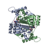
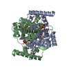
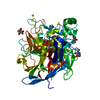
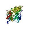
| Title | E. coli NfsA with nitrofurantoin | ||||||
 Components Components | Oxygen-insensitive NADPH nitroreductase | ||||||
 Keywords Keywords | OXIDOREDUCTASE / complex with Antibiotic substrate / flavoprotein / nitroreductase | ||||||
| Function / homology |  Function and homology information Function and homology informationNAD(P)H dehydrogenase (quinone) activity / Oxidoreductases / FMN binding / protein homodimerization activity / cytosol Similarity search - Function | ||||||
| Biological species |  | ||||||
| Method |  X-RAY DIFFRACTION / SYNCHROTRON / MOLECULAR REPLACEMENT / Resolution: 1.09 Å X-RAY DIFFRACTION / SYNCHROTRON / MOLECULAR REPLACEMENT / Resolution: 1.09 Å | ||||||
 Authors Authors | Day, M.D. / Jarrom, D. / Grainger, A.I. / Parr, R.J. / Hyde, E.I. / White, S.A. | ||||||
 Citation Citation | Journal: Biochem.J. / Year: 2021 Title: The structures of E. coli NfsA bound to the antibiotic nitrofurantoin; to 1,4-benzoquinone and to FMN. Authors: Day, M.A. / Jarrom, D. / Christofferson, A.J. / Graziano, A.E. / Anderson, J.L.R. / Searle, P.F. / Hyde, E.I. / White, S.A. | ||||||
| History |
|
- Structure visualization
| Structure viewer | Molecule: MolmilJmol/JSmol |
|---|
- Downloads & links
-Download
| PDBx/mmCIF format | 7nb9.cif.gz | 132.1 KB | Display | PDBx/mmCIF format |
|---|---|---|---|---|
| PDB format | pdb7nb9.ent.gz | 102.2 KB | Display | PDB format |
| PDBx/mmJSON format | 7nb9.json.gz | Tree view | PDBx/mmJSON format | |
| Others |  Other downloads Other downloads |
-Validation report
| Arichive directory | https://data.pdbj.org/pub/pdb/validation_reports/nb/7nb9ftp://data.pdbj.org/pub/pdb/validation_reports/nb/7nb9 | HTTPS FTP |
|---|
-Related structure data
| Related structure data |  7niyC  7nmpC  7nnxC  1f5vS S: Starting model for refinement C: citing same article ( |
|---|---|
| Similar structure data |


-Links
 PDBj
PDBj- Assembly
| Deposited unit | 
| |||||||||
|---|---|---|---|---|---|---|---|---|---|---|
| 1 | 
| |||||||||
| Unit cell |
| |||||||||
| Components on special symmetry positions |
|
-Components
| #1: Protein | Mass: 26832.664 Da / Num. of mol.: 1 Source method: isolated from a genetically manipulated source Source: (gene. exp.) Strain: K12 / Gene: nfsA, mda18, mdaA, ybjB, b0851, JW0835 / Plasmid: pPS1341A1 / Details (production host): pET24 derivative / Production host: |
|---|---|
| #2: Chemical | ChemComp-FMN /   Mass: 456.344 Da / Num. of mol.: 1 / Source method: obtained synthetically / Formula: C17H21N4O9P Mass: 456.344 Da / Num. of mol.: 1 / Source method: obtained synthetically / Formula: C17H21N4O9P |
| #3: Chemical | ChemComp-U6Z /   Mass: 238.157 Da / Num. of mol.: 1 / Source method: obtained synthetically / Formula: C8H6N4O5 / Feature type: SUBJECT OF INVESTIGATION / Comment: medication, antibiotic*YM Mass: 238.157 Da / Num. of mol.: 1 / Source method: obtained synthetically / Formula: C8H6N4O5 / Feature type: SUBJECT OF INVESTIGATION / Comment: medication, antibiotic*YM |
| #4: Chemical | ChemComp-DMS /   Mass: 78.133 Da / Num. of mol.: 1 / Source method: obtained synthetically / Formula: C2H6OS / Comment: DMSO, precipitant*YM Mass: 78.133 Da / Num. of mol.: 1 / Source method: obtained synthetically / Formula: C2H6OS / Comment: DMSO, precipitant*YM |
| #5: Water | ChemComp-HOH /  Mass: 18.015 Da / Num. of mol.: 297 / Source method: isolated from a natural source / Formula: H2O Mass: 18.015 Da / Num. of mol.: 297 / Source method: isolated from a natural source / Formula: H2O |
| Has ligand of interest | Y |
-Experimental details
-Experiment
| Experiment | Method: X-RAY DIFFRACTION / Number of used crystals: 1 |
|---|
- Sample preparation
| Crystal | Density Matthews: 2.05 Å3/Da / Density % sol: 39.94 % |
|---|---|
| Crystal grow | Temperature: 291 K / Method: vapor diffusion, sitting drop / pH: 7 Details: 100 mM imidazole pH 7, 20-26 % PEG 3,000, 3.5 mM nitrofurantoin, 20% DMSO |
-Data collection
| Diffraction | Mean temperature: 100 K / Serial crystal experiment: N |
|---|---|
| Diffraction source | Source: SYNCHROTRON / Site: ESRF  / Beamline: ID23-1 / Wavelength: 1 Å / Beamline: ID23-1 / Wavelength: 1 Å |
| Detector | Type: ADSC QUANTUM 315 / Detector: CCD / Date: Nov 21, 2010 |
| Radiation | Protocol: SINGLE WAVELENGTH / Monochromatic (M) / Laue (L): M / Scattering type: x-ray |
| Radiation wavelength | Wavelength: 1 Å / Relative weight: 1 |
| Reflection | Resolution: 1.09→45.69 Å / Num. obs: 83276 / % possible obs: 91.4 % / Redundancy: 3.4 % / Rsym value: 0.037 / Net I/σ(I): 19.4 |
| Reflection shell | Resolution: 1.09→1.15 Å / Redundancy: 2.33 % / Mean I/σ(I) obs: 3.69 / Num. unique obs: 9425 / Rsym value: 0.22 / % possible all: 64.5 |
- Processing
| Software |
| |||||||||||||||||||||||||||||||||||||||||||||||||||||||||||||||||||||||||||
|---|---|---|---|---|---|---|---|---|---|---|---|---|---|---|---|---|---|---|---|---|---|---|---|---|---|---|---|---|---|---|---|---|---|---|---|---|---|---|---|---|---|---|---|---|---|---|---|---|---|---|---|---|---|---|---|---|---|---|---|---|---|---|---|---|---|---|---|---|---|---|---|---|---|---|---|---|
| Refinement | Method to determine structure: MOLECULAR REPLACEMENT Starting model: 1F5V Resolution: 1.09→45.69 Å / Cor.coef. Fo:Fc: 0.983 / Cor.coef. Fo:Fc free: 0.979 / SU B: 0.837 / SU ML: 0.018 / Cross valid method: THROUGHOUT / σ(F): 0 / ESU R: 0.024 / ESU R Free: 0.025 / Stereochemistry target values: MAXIMUM LIKELIHOOD Details: HYDROGENS HAVE BEEN ADDED IN THE RIDING POSITIONS U VALUES : REFINED INDIVIDUALLY
| |||||||||||||||||||||||||||||||||||||||||||||||||||||||||||||||||||||||||||
| Solvent computation | Ion probe radii: 0.7 Å / Shrinkage radii: 0.7 Å / VDW probe radii: 1 Å / Solvent model: MASK | |||||||||||||||||||||||||||||||||||||||||||||||||||||||||||||||||||||||||||
| Displacement parameters | Biso max: 177.45 Å2 / Biso mean: 16.984 Å2 / Biso min: 7.11 Å2
| |||||||||||||||||||||||||||||||||||||||||||||||||||||||||||||||||||||||||||
| Refinement step | Cycle: final / Resolution: 1.09→45.69 Å
| |||||||||||||||||||||||||||||||||||||||||||||||||||||||||||||||||||||||||||
| Refine LS restraints |
| |||||||||||||||||||||||||||||||||||||||||||||||||||||||||||||||||||||||||||
| LS refinement shell | Resolution: 1.09→1.119 Å / Rfactor Rfree error: 0 / Total num. of bins used: 20
|
