 Movie
Movie Controller


+ Open data

- Basic information
| Entry | Database: PDB / ID: 7n9k | ||||||
|---|---|---|---|---|---|---|---|
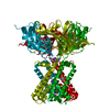
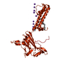
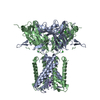
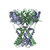
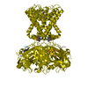
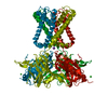
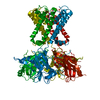
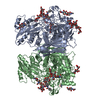
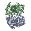
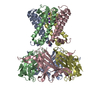
| Title | KirBac3.1 L124M mutant | ||||||
 Components Components | Inward rectifier potassium channel Kirbac3.1 | ||||||
 Keywords Keywords | MEMBRANE PROTEIN / potassium channel gating / mutant | ||||||
| Function / homology |  Function and homology information Function and homology informationinward rectifier potassium channel activity / regulation of monoatomic ion transmembrane transport / potassium ion import across plasma membrane / monoatomic ion channel complex / identical protein binding / plasma membrane Similarity search - Function | ||||||
| Biological species |  Magnetospirillum magnetotacticum (bacteria) Magnetospirillum magnetotacticum (bacteria) | ||||||
| Method |  X-RAY DIFFRACTION / SYNCHROTRON / MOLECULAR REPLACEMENT / molecular replacement / Resolution: 2.72 Å X-RAY DIFFRACTION / SYNCHROTRON / MOLECULAR REPLACEMENT / molecular replacement / Resolution: 2.72 Å | ||||||
 Authors Authors | Black, T.A. / Gulbis, J.M. | ||||||
 Citation Citation | Journal: Nat Commun / Year: 2022 Title: Ion currents through Kir potassium channels are gated by anionic lipids. Authors: Jin, R. / He, S. / Black, K.A. / Clarke, O.B. / Wu, D. / Bolla, J.R. / Johnson, P. / Periasamy, A. / Wardak, A. / Czabotar, P. / Colman, P.M. / Robinson, C.V. / Laver, D. / Smith, B.J. / Gulbis, J.M. | ||||||
| History |
|
- Structure visualization
| Structure viewer | Molecule: MolmilJmol/JSmol |
|---|
- Downloads & links
-Download
| PDBx/mmCIF format | 7n9k.cif.gz | 126.4 KB | Display | PDBx/mmCIF format |
|---|---|---|---|---|
| PDB format | pdb7n9k.ent.gz | 96.3 KB | Display | PDB format |
| PDBx/mmJSON format | 7n9k.json.gz | Tree view | PDBx/mmJSON format | |
| Others |  Other downloads Other downloads |
-Validation report
| Arichive directory | https://data.pdbj.org/pub/pdb/validation_reports/n9/7n9kftp://data.pdbj.org/pub/pdb/validation_reports/n9/7n9k | HTTPS FTP |
|---|
-Related structure data
| Related structure data |  7n9lC 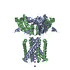 1xl4S S: Starting model for refinement C: citing same article ( |
|---|---|
| Similar structure data |
-Links
 PDBj
PDBj


- Assembly
| Deposited unit | 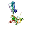
| ||||||||||||||||||
|---|---|---|---|---|---|---|---|---|---|---|---|---|---|---|---|---|---|---|---|
| 1 | 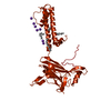
| ||||||||||||||||||
| Unit cell |
| ||||||||||||||||||
| Components on special symmetry positions |
|
-Components
-Protein , 1 types, 1 molecules A
| #1: Protein | Mass: 33768.676 Da / Num. of mol.: 1 / Mutation: C71S, L124M, C262S Source method: isolated from a genetically manipulated source Source: (gene. exp.) Magnetospirillum magnetotacticum (bacteria)Production host: |
|---|
-Non-polymers , 5 types, 52 molecules 


| #2: Chemical |  Mass: 787.121 Da / Num. of mol.: 3 / Source method: obtained synthetically / Formula: C44H85NO8P / Comment: DOPC, phospholipid*YM Mass: 787.121 Da / Num. of mol.: 3 / Source method: obtained synthetically / Formula: C44H85NO8P / Comment: DOPC, phospholipid*YM#3: Chemical | ChemComp-TMO / |  Mass: 75.110 Da / Num. of mol.: 1 / Source method: obtained synthetically / Formula: C3H9NO Mass: 75.110 Da / Num. of mol.: 1 / Source method: obtained synthetically / Formula: C3H9NO#4: Chemical | ChemComp-OCT / |  Mass: 114.229 Da / Num. of mol.: 1 / Source method: obtained synthetically / Formula: C8H18 Mass: 114.229 Da / Num. of mol.: 1 / Source method: obtained synthetically / Formula: C8H18#5: Chemical | ChemComp-K /  Mass: 39.098 Da / Num. of mol.: 7 / Source method: obtained synthetically / Formula: K Mass: 39.098 Da / Num. of mol.: 7 / Source method: obtained synthetically / Formula: K#6: Water | ChemComp-HOH / | Mass: 18.015 Da / Num. of mol.: 40 / Source method: isolated from a natural source / Formula: H2O |
|---|
-Details
| Has ligand of interest | N |
|---|
-Experimental details
-Experiment
| Experiment | Method: X-RAY DIFFRACTION / Number of used crystals: 1 |
|---|
- Sample preparation
| Crystal | Density Matthews: 3.77 Å3/Da / Density % sol: 67.35 % |
|---|---|
| Crystal grow | Temperature: 293 K / Method: vapor diffusion, sitting drop / pH: 7.5 Details: 90 mM HEPES pH 7.5, 2.5 w/v % PEG 8K, 2.5% w/v PEG 4K, 20.9% w/w PEG 400, 10% v/v glycerol. |
-Data collection
| Diffraction | Mean temperature: 100 K / Serial crystal experiment: N |
|---|---|
| Diffraction source | Source: SYNCHROTRON / Site: Australian Synchrotron  / Beamline: MX2 / Wavelength: 0.9537 Å / Beamline: MX2 / Wavelength: 0.9537 Å |
| Detector | Type: DECTRIS EIGER X 16M / Detector: PIXEL / Date: Feb 1, 2021 |
| Radiation | Protocol: SINGLE WAVELENGTH / Monochromatic (M) / Laue (L): M / Scattering type: x-ray |
| Radiation wavelength | Wavelength: 0.9537 Å / Relative weight: 1 |
| Reflection | Resolution: 2.71→47.663 Å / Num. obs: 14347 / % possible obs: 99.8 % / Redundancy: 7.22 % / Biso Wilson estimate: 61.17 Å2 / CC1/2: 1 / Rrim(I) all: 0.17 / Net I/σ(I): 10.52 |
| Reflection shell | Resolution: 2.71→2.89 Å / Mean I/σ(I) obs: 1.35 / Num. unique obs: 4231 / CC1/2: 0.579 / Rrim(I) all: 1.41 / % possible all: 99.2 |
-Phasing
| Phasing | Method: molecular replacement |
|---|
- Processing
| Software |
| ||||||||||||||||||||||||||||||||||||
|---|---|---|---|---|---|---|---|---|---|---|---|---|---|---|---|---|---|---|---|---|---|---|---|---|---|---|---|---|---|---|---|---|---|---|---|---|---|
| Refinement | Method to determine structure: MOLECULAR REPLACEMENT Starting model: 1xl4 Resolution: 2.72→42.08 Å / SU ML: 0.22 / Cross valid method: THROUGHOUT / σ(F): 1.36 / Phase error: 23.46 / Stereochemistry target values: ML
| ||||||||||||||||||||||||||||||||||||
| Solvent computation | Shrinkage radii: 0.9 Å / VDW probe radii: 1.11 Å / Solvent model: FLAT BULK SOLVENT MODEL | ||||||||||||||||||||||||||||||||||||
| Displacement parameters | Biso max: 166.24 Å2 / Biso mean: 67.2441 Å2 / Biso min: 26.71 Å2 | ||||||||||||||||||||||||||||||||||||
| Refinement step | Cycle: final / Resolution: 2.72→42.08 Å
| ||||||||||||||||||||||||||||||||||||
| LS refinement shell | Refine-ID: X-RAY DIFFRACTION / Rfactor Rfree error: 0 / Total num. of bins used: 5 / % reflection obs: 100 %
|
