 Movie
Movie Controller

[English] 日本語
 Yorodumi
- PDB-7ms1: Crystal structure of H28A mutant of Cg10062 with a covalent inter... -
+ Open data

- Basic information
| Entry | Database: PDB / ID: 7ms1 | ||||||
|---|---|---|---|---|---|---|---|
| Title | Crystal structure of H28A mutant of Cg10062 with a covalent intermediate of the hydration of acetylenecarboxylic acid | ||||||
 Components Components | 4-oxalocrotonate tautomerase | ||||||
 Keywords Keywords | HYDROLASE / tautomerase | ||||||
| Function / homology | Tautomerase, cis-CaaD-like / Putative oxalocrotonate tautomerase enzyme / Macrophage Migration Inhibitory Factor / Macrophage Migration Inhibitory Factor / Tautomerase/MIF superfamily / 2-Layer Sandwich / Alpha Beta / 3-HYDROXY-PROPANOIC ACID / 4-oxalocrotonate tautomerase Function and homology information Function and homology information | ||||||
| Biological species |  Corynebacterium glutamicum (bacteria) Corynebacterium glutamicum (bacteria) | ||||||
| Method |  X-RAY DIFFRACTION / SYNCHROTRON / MOLECULAR REPLACEMENT / Resolution: 2.95 Å X-RAY DIFFRACTION / SYNCHROTRON / MOLECULAR REPLACEMENT / Resolution: 2.95 Å | ||||||
 Authors Authors | Nayebi, G.H. / Geiger, J.H. / Draths, K. | ||||||
 Citation Citation | Journal: Biochemistry / Year: 2021 Title: Cg10062 Catalysis Forges a Link between Acetylenecarboxylic Acid and Bacterial Metabolism. Authors: Mathes Hewage, A. / Nayebi Gavgani, H. / Chi, D. / Qiu, B. / Geiger, J.H. / Draths, K. | ||||||
| History |
|
- Structure visualization
| Structure viewer | Molecule: MolmilJmol/JSmol |
|---|
- Downloads & links
-Download
| PDBx/mmCIF format | 7ms1.cif.gz | 77.2 KB | Display | PDBx/mmCIF format |
|---|---|---|---|---|
| PDB format | pdb7ms1.ent.gz | 55.5 KB | Display | PDB format |
| PDBx/mmJSON format | 7ms1.json.gz | Tree view | PDBx/mmJSON format | |
| Others |  Other downloads Other downloads |
-Validation report
| Arichive directory | https://data.pdbj.org/pub/pdb/validation_reports/ms/7ms1ftp://data.pdbj.org/pub/pdb/validation_reports/ms/7ms1 | HTTPS FTP |
|---|
-Related structure data
| Related structure data | 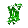 7ms0SC 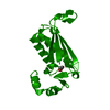 7ms3C 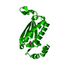 7ms8C 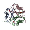 7ms9C S: Starting model for refinement C: citing same article ( |
|---|---|
| Similar structure data |


-Links
 PDBj
PDBj- Assembly
| Deposited unit | 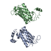
| ||||||||||||
|---|---|---|---|---|---|---|---|---|---|---|---|---|---|
| 1 | 
| ||||||||||||
| 2 | 
| ||||||||||||
| Unit cell |
|
-Components
| #1: Protein | Mass: 18971.182 Da / Num. of mol.: 2 / Mutation: H28A Source method: isolated from a genetically manipulated source Source: (gene. exp.) Corynebacterium glutamicum (bacteria)Gene: APT58_00490, AUO95_07180, CS176_0056, FM102_14895, KaCgl_17770, KbCgl_30240 Production host: #2: Chemical | ChemComp-3OH / |   Mass: 90.078 Da / Num. of mol.: 1 / Source method: obtained synthetically / Formula: C3H6O3 / Feature type: SUBJECT OF INVESTIGATION Mass: 90.078 Da / Num. of mol.: 1 / Source method: obtained synthetically / Formula: C3H6O3 / Feature type: SUBJECT OF INVESTIGATION#3: Water | ChemComp-HOH / |  Mass: 18.015 Da / Num. of mol.: 1 / Source method: isolated from a natural source / Formula: H2O Mass: 18.015 Da / Num. of mol.: 1 / Source method: isolated from a natural source / Formula: H2OHas ligand of interest | Y | Has protein modification | Y | |
|---|
-Experimental details
-Experiment
| Experiment | Method: X-RAY DIFFRACTION / Number of used crystals: 1 |
|---|
- Sample preparation
| Crystal | Density Matthews: 3.54 Å3/Da / Density % sol: 65.3 % |
|---|---|
| Crystal grow | Temperature: 300 K / Method: vapor diffusion, hanging drop / pH: 5.6 Details: 50 mM ammonium acetate, 25 mM sodium citrate tribasic dihydrate, pH 5.6, 7.5% w/v PEG4000 |
-Data collection
| Diffraction | Mean temperature: 100 K / Serial crystal experiment: N |
|---|---|
| Diffraction source | Source: SYNCHROTRON / Site: APS  / Beamline: 21-ID-D / Wavelength: 1 Å / Beamline: 21-ID-D / Wavelength: 1 Å |
| Detector | Type: DECTRIS EIGER X 9M / Detector: PIXEL / Date: Jun 2, 2019 |
| Radiation | Protocol: SINGLE WAVELENGTH / Monochromatic (M) / Laue (L): M / Scattering type: x-ray |
| Radiation wavelength | Wavelength: 1 Å / Relative weight: 1 |
| Reflection | Resolution: 2.95→35.42 Å / Num. obs: 11335 / % possible obs: 96.45 % / Redundancy: 5.9 % / Rmerge(I) obs: 0.163 / Rpim(I) all: 0.071 / Rrim(I) all: 0.179 / Net I/σ(I): 11.3 |
| Reflection shell | Resolution: 2.95→3.06 Å / Redundancy: 5.7 % / Rmerge(I) obs: 1 / Num. unique obs: 1112 / Χ2: 0.516 |
- Processing
| Software |
| ||||||||||||||||||||||||
|---|---|---|---|---|---|---|---|---|---|---|---|---|---|---|---|---|---|---|---|---|---|---|---|---|---|
| Refinement | Method to determine structure: MOLECULAR REPLACEMENT Starting model: PDB entry 7MS0 Resolution: 2.95→35.42 Å / Cross valid method: FREE R-VALUE Stereochemistry target values: GeoStd + Monomer Library + CDL v1.2
| ||||||||||||||||||||||||
| Refinement step | Cycle: LAST / Resolution: 2.95→35.42 Å
| ||||||||||||||||||||||||
| Refine LS restraints |
|
