 Movie
Movie Controller


+ Open data

- Basic information
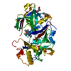
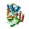
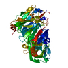
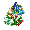
| Entry | Database: PDB / ID: 7mq7 | ||||||
|---|---|---|---|---|---|---|---|
| Title | Tetragonal Maltose Binding Protein | ||||||
 Components Components | Maltodextrin-binding protein | ||||||
 Keywords Keywords | SUGAR BINDING PROTEIN / Maltose Binding Protein | ||||||
| Function / homology | beta-maltose / :  Function and homology information Function and homology information | ||||||
| Biological species |  | ||||||
| Method |  X-RAY DIFFRACTION / SYNCHROTRON / MOLECULAR REPLACEMENT / Resolution: 1.95 Å X-RAY DIFFRACTION / SYNCHROTRON / MOLECULAR REPLACEMENT / Resolution: 1.95 Å | ||||||
 Authors Authors | Thaker, A. / Sirajudeen, L. / Simmons, C.R. / Nannenga, B.L. | ||||||
| Funding support |  United States, 1items United States, 1items
| ||||||
 Citation Citation | Journal: Biotechnol.Bioeng. / Year: 2021 Title: Structure-guided identification of a peptide for bio-enabled gold nanoparticle synthesis. Authors: Thaker, A. / Sirajudeen, L. / Simmons, C.R. / Nannenga, B.L. | ||||||
| History |
|
- Structure visualization
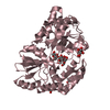
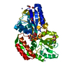
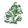
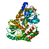
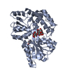
| Structure viewer | Molecule: MolmilJmol/JSmol |
|---|
- Downloads & links
-Download
| PDBx/mmCIF format | 7mq7.cif.gz | 175.3 KB | Display | PDBx/mmCIF format |
|---|---|---|---|---|
| PDB format | pdb7mq7.ent.gz | 136.9 KB | Display | PDB format |
| PDBx/mmJSON format | 7mq7.json.gz | Tree view | PDBx/mmJSON format | |
| Others |  Other downloads Other downloads |
-Validation report
| Arichive directory | https://data.pdbj.org/pub/pdb/validation_reports/mq/7mq7ftp://data.pdbj.org/pub/pdb/validation_reports/mq/7mq7 | HTTPS FTP |
|---|
-Related structure data
| Related structure data |  7mq6C  1anfS C: citing same article ( S: Starting model for refinement |
|---|---|
| Similar structure data |


-Links
 PDBj
PDBj- Assembly
| Deposited unit | 
| ||||||||
|---|---|---|---|---|---|---|---|---|---|
| 1 |
| ||||||||
| Unit cell |
|
-Components
| #1: Protein | Mass: 41084.586 Da / Num. of mol.: 1 Source method: isolated from a genetically manipulated source Source: (gene. exp.) | ||||||
|---|---|---|---|---|---|---|---|
| #2: Polysaccharide | alpha-D-glucopyranose-(1-4)-beta-D-glucopyranose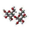  Source method: isolated from a genetically manipulated source Details: oligosaccharide / References: beta-maltose | ||||||
| #3: Chemical |   Mass: 96.063 Da / Num. of mol.: 3 / Source method: obtained synthetically / Formula: SO4 Mass: 96.063 Da / Num. of mol.: 3 / Source method: obtained synthetically / Formula: SO4#4: Chemical |   Mass: 35.453 Da / Num. of mol.: 2 / Source method: obtained synthetically / Formula: Cl Mass: 35.453 Da / Num. of mol.: 2 / Source method: obtained synthetically / Formula: Cl#5: Water | ChemComp-HOH / |  Mass: 18.015 Da / Num. of mol.: 362 / Source method: isolated from a natural source / Formula: H2O Mass: 18.015 Da / Num. of mol.: 362 / Source method: isolated from a natural source / Formula: H2OHas ligand of interest | N | |
-Experimental details
-Experiment
| Experiment | Method: X-RAY DIFFRACTION / Number of used crystals: 1 |
|---|
- Sample preparation
| Crystal | Density Matthews: 3.07 Å3/Da / Density % sol: 59.99 % |
|---|---|
| Crystal grow | Temperature: 298 K / Method: vapor diffusion Details: 2.4 M ammonium sulfate, 0.1 M sodium acetate pH 4.5 |
-Data collection
| Diffraction | Mean temperature: 77 K / Serial crystal experiment: N |
|---|---|
| Diffraction source | Source: SYNCHROTRON / Site: APS / Beamline: 19-ID / Wavelength: 1.08724 Å |
| Detector | Type: DECTRIS PILATUS3 6M / Detector: PIXEL / Date: Mar 21, 2017 |
| Radiation | Protocol: SINGLE WAVELENGTH / Monochromatic (M) / Laue (L): M / Scattering type: x-ray |
| Radiation wavelength | Wavelength: 1.08724 Å / Relative weight: 1 |
| Reflection | Resolution: 1.95→32.001 Å / Num. obs: 35971 / % possible obs: 95 % / Redundancy: 8.3 % / Biso Wilson estimate: 29.08 Å2 / CC1/2: 0.992 / Net I/σ(I): 16 |
| Reflection shell | Resolution: 1.95→2.01 Å / Num. unique obs: 3070 / CC1/2: 0.948 |
- Processing
| Software |
| ||||||||||||||||||||||||||||||||||||||||||||||||||||||||||||||||||||||||||||||
|---|---|---|---|---|---|---|---|---|---|---|---|---|---|---|---|---|---|---|---|---|---|---|---|---|---|---|---|---|---|---|---|---|---|---|---|---|---|---|---|---|---|---|---|---|---|---|---|---|---|---|---|---|---|---|---|---|---|---|---|---|---|---|---|---|---|---|---|---|---|---|---|---|---|---|---|---|---|---|---|
| Refinement | Method to determine structure: MOLECULAR REPLACEMENT Starting model: 1anf Resolution: 1.95→32.001 Å / SU ML: 0.21 / Cross valid method: FREE R-VALUE / σ(F): 1.33 / Phase error: 26.23 / Stereochemistry target values: ML
| ||||||||||||||||||||||||||||||||||||||||||||||||||||||||||||||||||||||||||||||
| Solvent computation | Shrinkage radii: 0.9 Å / VDW probe radii: 1.11 Å / Solvent model: FLAT BULK SOLVENT MODEL | ||||||||||||||||||||||||||||||||||||||||||||||||||||||||||||||||||||||||||||||
| Displacement parameters | Biso max: 113.36 Å2 / Biso mean: 40.7854 Å2 / Biso min: 26.28 Å2 | ||||||||||||||||||||||||||||||||||||||||||||||||||||||||||||||||||||||||||||||
| Refinement step | Cycle: final / Resolution: 1.95→32.001 Å
| ||||||||||||||||||||||||||||||||||||||||||||||||||||||||||||||||||||||||||||||
| LS refinement shell | Refine-ID: X-RAY DIFFRACTION / Rfactor Rfree error: 0
| ||||||||||||||||||||||||||||||||||||||||||||||||||||||||||||||||||||||||||||||
| Refinement TLS params. | Method: refined / Origin x: 22.6173 Å / Origin y: 63.1743 Å / Origin z: 0.4661 Å
| ||||||||||||||||||||||||||||||||||||||||||||||||||||||||||||||||||||||||||||||
| Refinement TLS group |
|
