 Movie
Movie Controller

[English] 日本語
 Yorodumi
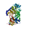
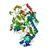
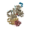
- PDB-7ml5: Structure of the Starch Branching Enzyme I (BEI) complexed with m... -
+ Open data

- Basic information
| Entry | Database: PDB / ID: 7ml5 | ||||||
|---|---|---|---|---|---|---|---|
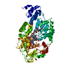
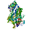
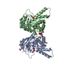
| Title | Structure of the Starch Branching Enzyme I (BEI) complexed with maltododecaose from Oryza sativa L | ||||||
 Components Components | Isoform 2 of 1,4-alpha-glucan-branching enzyme, chloroplastic/amyloplastic | ||||||
 Keywords Keywords | TRANSFERASE/SUBSTRATE / rBEI / maltododecaose / Branching Enzyme I / TRANSFERASE / TRANSFERASE-SUBSTRATE complex | ||||||
| Function / homology |  Function and homology information Function and homology informationstarch metabolic process / amyloplast / cation binding / starch biosynthetic process / 1,4-alpha-glucan branching enzyme / 1,4-alpha-glucan branching enzyme activity / starch catabolic process / glycogen biosynthetic process / hydrolase activity, hydrolyzing O-glycosyl compounds / chloroplast ...starch metabolic process / amyloplast / cation binding / starch biosynthetic process / 1,4-alpha-glucan branching enzyme / 1,4-alpha-glucan branching enzyme activity / starch catabolic process / glycogen biosynthetic process / hydrolase activity, hydrolyzing O-glycosyl compounds / chloroplast / carbohydrate metabolic process / cytoplasm Similarity search - Function | ||||||
| Biological species |  | ||||||
| Method |  X-RAY DIFFRACTION / SYNCHROTRON / MOLECULAR REPLACEMENT / Resolution: 2.35 Å X-RAY DIFFRACTION / SYNCHROTRON / MOLECULAR REPLACEMENT / Resolution: 2.35 Å | ||||||
 Authors Authors | Nayebi Gavgani, H. / Fawaz, R. / Geiger, J.H. | ||||||
| Funding support |  United States, 1items United States, 1items
| ||||||
 Citation Citation | Journal: J.Biol.Chem. / Year: 2021 Title: A structural explanation for the mechanism and specificity of plant branching enzymes I and IIb. Authors: Gavgani, H.N. / Fawaz, R. / Ehyaei, N. / Walls, D. / Pawlowski, K. / Fulgos, R. / Park, S. / Assar, Z. / Ghanbarpour, A. / Geiger, J.H. | ||||||
| History |
|
- Structure visualization
| Structure viewer | Molecule: MolmilJmol/JSmol |
|---|
- Downloads & links
-Download
| PDBx/mmCIF format | 7ml5.cif.gz | 181.2 KB | Display | PDBx/mmCIF format |
|---|---|---|---|---|
| PDB format | pdb7ml5.ent.gz | 128.8 KB | Display | PDB format |
| PDBx/mmJSON format | 7ml5.json.gz | Tree view | PDBx/mmJSON format | |
| Others |  Other downloads Other downloads |
-Validation report
| Arichive directory | https://data.pdbj.org/pub/pdb/validation_reports/ml/7ml5ftp://data.pdbj.org/pub/pdb/validation_reports/ml/7ml5 | HTTPS FTP |
|---|
-Related structure data
| Related structure data | 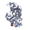 3vu2S S: Starting model for refinement |
|---|---|
| Similar structure data |


-Links
 PDBj
PDBj

- Assembly
| Deposited unit | 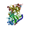
| ||||||||||||
|---|---|---|---|---|---|---|---|---|---|---|---|---|---|
| 1 |
| ||||||||||||
| Unit cell |
|
-Components
| #1: Protein | Mass: 81101.477 Da / Num. of mol.: 1 / Mutation: L40M, V280M, S443P, T669A Source method: isolated from a genetically manipulated source Details: L40M-V280M-S443P-T669A is the protein variant that produced the crystal. Source: (gene. exp.) Gene: SBE1, RBE1, Os06g0726400, LOC_Os06g51084, P0017G10.8-1, P0017G10.8-2, P0548E04.28-1, P0548E04.28-2 Production host:  References: UniProt: Q01401, 1,4-alpha-glucan branching enzyme |
|---|---|
| #2: Polysaccharide | alpha-D-glucopyranose-(1-4)-alpha-D-glucopyranose-(1-4)-alpha-D-glucopyranose-(1-4)-alpha-D- ...alpha-D-glucopyranose-(1-4)-alpha-D-glucopyranose-(1-4)-alpha-D-glucopyranose-(1-4)-alpha-D-glucopyranose-(1-4)-alpha-D-glucopyranose-(1-4)-alpha-D-glucopyranose-(1-4)-alpha-D-glucopyranose-(1-4)-alpha-D-glucopyranose-(1-4)-alpha-D-glucopyranose-(1-4)-alpha-D-glucopyranose-(1-4)-alpha-D-glucopyranose-(1-4)-alpha-D-glucopyranose Type: oligosaccharide / Mass: 1963.705 Da / Num. of mol.: 1 / Source method: isolated from a natural source |
| #3: Polysaccharide | alpha-D-glucopyranose-(1-4)-alpha-D-glucopyranose-(1-4)-alpha-D-glucopyranose-(1-4)-alpha-D-glucopyranose |
| #4: Water | ChemComp-HOH /  Mass: 18.015 Da / Num. of mol.: 198 / Source method: isolated from a natural source / Formula: H2O Mass: 18.015 Da / Num. of mol.: 198 / Source method: isolated from a natural source / Formula: H2O |
| Has ligand of interest | Y |
-Experimental details
-Experiment
| Experiment | Method: X-RAY DIFFRACTION / Number of used crystals: 1 |
|---|
- Sample preparation
| Crystal | Density Matthews: 2.01 Å3/Da / Density % sol: 38.83 % |
|---|---|
| Crystal grow | Temperature: 300 K / Method: vapor diffusion, hanging drop / pH: 6.9 Details: 30% PEG8K, 550 mM sodium acetate, and 100 mM sodium cacodylate (pH 6.9) |
-Data collection
| Diffraction | Mean temperature: 100 K / Serial crystal experiment: N |
|---|---|
| Diffraction source | Source: SYNCHROTRON / Site: SPring-8  / Beamline: BL38B1 / Wavelength: 1 Å / Beamline: BL38B1 / Wavelength: 1 Å |
| Detector | Type: ADSC QUANTUM 315 / Detector: CCD / Date: Feb 2, 2010 |
| Radiation | Protocol: SINGLE WAVELENGTH / Monochromatic (M) / Laue (L): M / Scattering type: x-ray |
| Radiation wavelength | Wavelength: 1 Å / Relative weight: 1 |
| Reflection | Resolution: 2.35→39.97 Å / Num. obs: 28616 / % possible obs: 95.24 % / Redundancy: 4.1 % / Biso Wilson estimate: 36.88 Å2 / Rmerge(I) obs: 0.091 / Net I/σ(I): 14.7 |
| Reflection shell | Resolution: 2.35→2.434 Å / Rmerge(I) obs: 0.316 / Num. unique obs: 2155 / % possible all: 73.45 |
- Processing
| Software |
| |||||||||||||||||||||||||||||||||||||||||||||||||||||||||||||||||||||||||||||
|---|---|---|---|---|---|---|---|---|---|---|---|---|---|---|---|---|---|---|---|---|---|---|---|---|---|---|---|---|---|---|---|---|---|---|---|---|---|---|---|---|---|---|---|---|---|---|---|---|---|---|---|---|---|---|---|---|---|---|---|---|---|---|---|---|---|---|---|---|---|---|---|---|---|---|---|---|---|---|
| Refinement | Method to determine structure: MOLECULAR REPLACEMENT Starting model: 3VU2 Resolution: 2.35→39.97 Å / SU ML: 0.2826 / Cross valid method: FREE R-VALUE / σ(F): 1.37 / Phase error: 23.5375 Stereochemistry target values: GeoStd + Monomer Library + CDL v1.2
| |||||||||||||||||||||||||||||||||||||||||||||||||||||||||||||||||||||||||||||
| Solvent computation | Shrinkage radii: 0.9 Å / VDW probe radii: 1.11 Å / Solvent model: FLAT BULK SOLVENT MODEL | |||||||||||||||||||||||||||||||||||||||||||||||||||||||||||||||||||||||||||||
| Displacement parameters | Biso mean: 37.32 Å2 | |||||||||||||||||||||||||||||||||||||||||||||||||||||||||||||||||||||||||||||
| Refinement step | Cycle: LAST / Resolution: 2.35→39.97 Å
| |||||||||||||||||||||||||||||||||||||||||||||||||||||||||||||||||||||||||||||
| Refine LS restraints |
| |||||||||||||||||||||||||||||||||||||||||||||||||||||||||||||||||||||||||||||
| LS refinement shell |
|
