 Movie
Movie Controller

[English] 日本語
 Yorodumi
- PDB-7mjf: Crystal structure of Candidatus Liberibacter solanacearum dihydro... -
+ Open data

- Basic information
| Entry | Database: PDB / ID: 7mjf | ||||||
|---|---|---|---|---|---|---|---|
| Title | Crystal structure of Candidatus Liberibacter solanacearum dihydrodipicolinate synthase with pyruvate and succinic semi-aldehyde bound in active site | ||||||
 Components Components | 4-hydroxy-tetrahydrodipicolinate synthase | ||||||
 Keywords Keywords | LYASE / dihydrodipicolinate / Liberibacter | ||||||
| Function / homology |  Function and homology information Function and homology information4-hydroxy-tetrahydrodipicolinate synthase / 4-hydroxy-tetrahydrodipicolinate synthase activity / : / : / cytosol Similarity search - Function | ||||||
| Biological species |  Candidatus Liberibacter solanacearum (bacteria) Candidatus Liberibacter solanacearum (bacteria) | ||||||
| Method |  X-RAY DIFFRACTION / SYNCHROTRON / MOLECULAR REPLACEMENT / Resolution: 2.2 Å X-RAY DIFFRACTION / SYNCHROTRON / MOLECULAR REPLACEMENT / Resolution: 2.2 Å | ||||||
 Authors Authors | Gilkes, J. / Frampton, R.A. / Board, A.J. / Sheen, C.R. / Smith, G.R. / Dobson, R.C.J. | ||||||
 Citation Citation | Journal: To Be Published Title: Crystal structure of Candidatus Liberibacter solanacearum dihydrodipicolinate synthase with pyruvate and succinic semi-aldehyde bound in active site Authors: Gilkes, J. / Frampton, R.A. / Board, A.J. / Sheen, C.R. / Smith, G.R. / Dobson, R.C.J. | ||||||
| History |
|
- Structure visualization
| Structure viewer | Molecule: MolmilJmol/JSmol |
|---|
- Downloads & links
-Download
| PDBx/mmCIF format | 7mjf.cif.gz | 701.9 KB | Display | PDBx/mmCIF format |
|---|---|---|---|---|
| PDB format | pdb7mjf.ent.gz | 587.1 KB | Display | PDB format |
| PDBx/mmJSON format | 7mjf.json.gz | Tree view | PDBx/mmJSON format | |
| Others |  Other downloads Other downloads |
-Validation report
| Arichive directory | https://data.pdbj.org/pub/pdb/validation_reports/mj/7mjfftp://data.pdbj.org/pub/pdb/validation_reports/mj/7mjf | HTTPS FTP |
|---|
-Related structure data
| Related structure data |  7loyS S: Starting model for refinement |
|---|---|
| Similar structure data |

-Links
 PDBj
PDBj- Assembly
| Deposited unit | 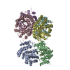
| ||||||||
|---|---|---|---|---|---|---|---|---|---|
| 1 | 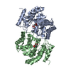
| ||||||||
| 2 | 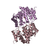
| ||||||||
| 3 | 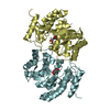
| ||||||||
| Unit cell |
|
-Components
| #1: Protein | Mass: 31867.117 Da / Num. of mol.: 6 Source method: isolated from a genetically manipulated source Source: (gene. exp.) Candidatus Liberibacter solanacearum (bacteria)Gene: dapA, DJ66_0589 / Production host: References: UniProt: A0A0F4VK59, 4-hydroxy-tetrahydrodipicolinate synthase #2: Chemical | ChemComp-E8U / (   Mass: 190.151 Da / Num. of mol.: 6 Mass: 190.151 Da / Num. of mol.: 6Source method: isolated from a genetically manipulated source Formula: C7H10O6 / Details: Succinic acid in 2 stereochemistry conformations / References: 4-hydroxy-tetrahydrodipicolinate synthase #3: Chemical | ChemComp-ZGM / (   Mass: 190.151 Da / Num. of mol.: 6 Mass: 190.151 Da / Num. of mol.: 6Source method: isolated from a genetically manipulated source Formula: C7H10O6 #4: Water | ChemComp-HOH / |  Mass: 18.015 Da / Num. of mol.: 897 / Source method: isolated from a natural source / Formula: H2O Mass: 18.015 Da / Num. of mol.: 897 / Source method: isolated from a natural source / Formula: H2OHas ligand of interest | Y | Has protein modification | Y | |
|---|
-Experimental details
-Experiment
| Experiment | Method: X-RAY DIFFRACTION / Number of used crystals: 1 |
|---|
- Sample preparation
| Crystal | Density Matthews: 2.69 Å3/Da / Density % sol: 54.34 % |
|---|---|
| Crystal grow | Temperature: 293 K / Method: vapor diffusion, sitting drop / Details: 20% w/v PEG3000, 0.1 M sodium citrate, pH 5.5 |
-Data collection
| Diffraction | Mean temperature: 110 K / Serial crystal experiment: N |
|---|---|
| Diffraction source | Source: SYNCHROTRON / Site: Australian Synchrotron  / Beamline: MX2 / Wavelength: 0.953723 Å / Beamline: MX2 / Wavelength: 0.953723 Å |
| Detector | Type: DECTRIS EIGER X 16M / Detector: PIXEL / Date: Dec 7, 2018 |
| Radiation | Protocol: SINGLE WAVELENGTH / Monochromatic (M) / Laue (L): M / Scattering type: x-ray |
| Radiation wavelength | Wavelength: 0.953723 Å / Relative weight: 1 |
| Reflection | Resolution: 1.925→45.812 Å / Num. obs: 195500 / % possible obs: 99.5 % / Redundancy: 3.9 % / Net I/σ(I): 8.32 |
| Reflection shell | Resolution: 1.93→1.99 Å |
- Processing
| Software |
| ||||||||||||||||||||||||||||||||||||||||||||||||||||||||||||||||||||||||||||||||||||||||||||||||||||||||||||||||||||||||||||||||||||||||||||||||||||||||||||||||||||||||||||||||||||||||||||||||||||||||||||||||||
|---|---|---|---|---|---|---|---|---|---|---|---|---|---|---|---|---|---|---|---|---|---|---|---|---|---|---|---|---|---|---|---|---|---|---|---|---|---|---|---|---|---|---|---|---|---|---|---|---|---|---|---|---|---|---|---|---|---|---|---|---|---|---|---|---|---|---|---|---|---|---|---|---|---|---|---|---|---|---|---|---|---|---|---|---|---|---|---|---|---|---|---|---|---|---|---|---|---|---|---|---|---|---|---|---|---|---|---|---|---|---|---|---|---|---|---|---|---|---|---|---|---|---|---|---|---|---|---|---|---|---|---|---|---|---|---|---|---|---|---|---|---|---|---|---|---|---|---|---|---|---|---|---|---|---|---|---|---|---|---|---|---|---|---|---|---|---|---|---|---|---|---|---|---|---|---|---|---|---|---|---|---|---|---|---|---|---|---|---|---|---|---|---|---|---|---|---|---|---|---|---|---|---|---|---|---|---|---|---|---|---|---|
| Refinement | Method to determine structure: MOLECULAR REPLACEMENT Starting model: PDB ENTRY 7LOY Resolution: 2.2→45.1 Å / SU ML: 0.26 / Cross valid method: THROUGHOUT / σ(F): 1.35 / Phase error: 22.89 / Stereochemistry target values: ML
| ||||||||||||||||||||||||||||||||||||||||||||||||||||||||||||||||||||||||||||||||||||||||||||||||||||||||||||||||||||||||||||||||||||||||||||||||||||||||||||||||||||||||||||||||||||||||||||||||||||||||||||||||||
| Solvent computation | Shrinkage radii: 0.9 Å / VDW probe radii: 1.11 Å / Solvent model: FLAT BULK SOLVENT MODEL | ||||||||||||||||||||||||||||||||||||||||||||||||||||||||||||||||||||||||||||||||||||||||||||||||||||||||||||||||||||||||||||||||||||||||||||||||||||||||||||||||||||||||||||||||||||||||||||||||||||||||||||||||||
| Refinement step | Cycle: LAST / Resolution: 2.2→45.1 Å
| ||||||||||||||||||||||||||||||||||||||||||||||||||||||||||||||||||||||||||||||||||||||||||||||||||||||||||||||||||||||||||||||||||||||||||||||||||||||||||||||||||||||||||||||||||||||||||||||||||||||||||||||||||
| Refine LS restraints |
| ||||||||||||||||||||||||||||||||||||||||||||||||||||||||||||||||||||||||||||||||||||||||||||||||||||||||||||||||||||||||||||||||||||||||||||||||||||||||||||||||||||||||||||||||||||||||||||||||||||||||||||||||||
| LS refinement shell |
| ||||||||||||||||||||||||||||||||||||||||||||||||||||||||||||||||||||||||||||||||||||||||||||||||||||||||||||||||||||||||||||||||||||||||||||||||||||||||||||||||||||||||||||||||||||||||||||||||||||||||||||||||||
| Refinement TLS params. | Method: refined / Origin x: 17.4676 Å / Origin y: -2.3157 Å / Origin z: 42.7004 Å
| ||||||||||||||||||||||||||||||||||||||||||||||||||||||||||||||||||||||||||||||||||||||||||||||||||||||||||||||||||||||||||||||||||||||||||||||||||||||||||||||||||||||||||||||||||||||||||||||||||||||||||||||||||
| Refinement TLS group | Selection details: ALL |
