 Movie
Movie Controller

[English] 日本語
 Yorodumi
- PDB-5t25: Kinetic, Spectral and Structural Characterization of the Slow Bin... -
+ Open data

- Basic information
| Entry | Database: PDB / ID: 5t25 | ||||||
|---|---|---|---|---|---|---|---|
| Title | Kinetic, Spectral and Structural Characterization of the Slow Binding Inhibitor Acetopyruvate with Dihydrodipicolinate Synthase from Escherichia coli. | ||||||
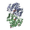
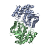
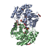
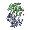
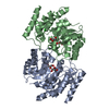
 Components Components | 4-hydroxy-tetrahydrodipicolinate synthase | ||||||
 Keywords Keywords | HYDROLASE / Dihydrodipicolinate Synthase / Kinetics / Acetopyruvate modification | ||||||
| Function / homology |  Function and homology information Function and homology information4-hydroxy-tetrahydrodipicolinate synthase / 4-hydroxy-tetrahydrodipicolinate synthase activity / : / : / cytosol Similarity search - Function | ||||||
| Biological species |  | ||||||
| Method |  X-RAY DIFFRACTION / MOLECULAR REPLACEMENT / Resolution: 1.991 Å X-RAY DIFFRACTION / MOLECULAR REPLACEMENT / Resolution: 1.991 Å | ||||||
 Authors Authors | Chooback, L. / Thomas, L.M. / Karsten, W.E. / Fleming, C.D. / Seabourn, P. | ||||||
| Funding support |  United States, 1items United States, 1items
| ||||||
 Citation Citation | Journal: To Be Published Title: Kinetic, Spectral and Structural Characterization of the Slow Binding Inhibitor Acetopyruvate with Dihydrodipicolinate Synthase from Escherichia coli. Authors: Chooback, L. / Thomas, L.M. / Karsten, W.E. / Fleming, C.D. / Seabourn, P. | ||||||
| History |
|
- Structure visualization
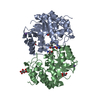
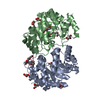
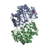
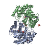
| Structure viewer | Molecule: MolmilJmol/JSmol |
|---|
- Downloads & links
-Download
| PDBx/mmCIF format | 5t25.cif.gz | 139.6 KB | Display | PDBx/mmCIF format |
|---|---|---|---|---|
| PDB format | pdb5t25.ent.gz | 106.5 KB | Display | PDB format |
| PDBx/mmJSON format | 5t25.json.gz | Tree view | PDBx/mmJSON format | |
| Others |  Other downloads Other downloads |
-Validation report
| Arichive directory | https://data.pdbj.org/pub/pdb/validation_reports/t2/5t25ftp://data.pdbj.org/pub/pdb/validation_reports/t2/5t25 | HTTPS FTP |
|---|
-Related structure data
| Related structure data |  5t26C  1yxcS C: citing same article ( S: Starting model for refinement |
|---|---|
| Similar structure data |


-Links
 PDBj
PDBj- Assembly
| Deposited unit | 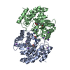
| ||||||||
|---|---|---|---|---|---|---|---|---|---|
| 1 |
| ||||||||
| Unit cell |
|
-Components
| #1: Protein | Mass: 31554.160 Da / Num. of mol.: 2 Source method: isolated from a genetically manipulated source Source: (gene. exp.) Strain: IAI1 / Gene: dapA, ECIAI1_2529 / Production host: References: UniProt: B7M7I1, 4-hydroxy-tetrahydrodipicolinate synthase #2: Chemical |   Type: L-peptide linking / Mass: 147.195 Da / Num. of mol.: 2 / Source method: obtained synthetically / Formula: C6H15N2O2 Type: L-peptide linking / Mass: 147.195 Da / Num. of mol.: 2 / Source method: obtained synthetically / Formula: C6H15N2O2#3: Chemical | ChemComp-NA /   Mass: 22.990 Da / Num. of mol.: 10 / Source method: obtained synthetically / Formula: Na Mass: 22.990 Da / Num. of mol.: 10 / Source method: obtained synthetically / Formula: Na#4: Water | ChemComp-HOH / |  Mass: 18.015 Da / Num. of mol.: 660 / Source method: isolated from a natural source / Formula: H2O Mass: 18.015 Da / Num. of mol.: 660 / Source method: isolated from a natural source / Formula: H2O |
|---|
-Experimental details
-Experiment
| Experiment | Method: X-RAY DIFFRACTION / Number of used crystals: 1 |
|---|
- Sample preparation
| Crystal | Density Matthews: 2.43 Å3/Da / Density % sol: 49.45 % |
|---|---|
| Crystal grow | Temperature: 293 K / Method: vapor diffusion, hanging drop / pH: 7.1 Details: 20% PEG3350 200mM Sodium Tartarate 14.7 mM HEPES pH 7.1 |
-Data collection
| Diffraction | Mean temperature: 100 K |
|---|---|
| Diffraction source | Source: ROTATING ANODE / Type: RIGAKU MICROMAX-007 HF / Wavelength: 1.54178 Å |
| Detector | Type: DECTRIS PILATUS 200K / Detector: PIXEL / Date: Jul 10, 2015 / Details: VeriMaxHF |
| Radiation | Monochromator: Multilayer optics / Protocol: SINGLE WAVELENGTH / Monochromatic (M) / Laue (L): M / Scattering type: x-ray |
| Radiation wavelength | Wavelength: 1.54178 Å / Relative weight: 1 |
| Reflection | Resolution: 2→81 Å / Num. obs: 193325 / % possible obs: 100 % / Redundancy: 4.5 % / Net I/σ(I): 9.8 |
| Reflection shell | Resolution: 2→2.03 Å / Redundancy: 3.1 % / Rmerge(I) obs: 0.54 / Mean I/σ(I) obs: 1.911 / CC1/2: 0.809 / % possible all: 100 |
- Processing
| Software |
| ||||||||||||||||||||||||||||||||||||||||||||||||||||||||||||||||||||||||||||||||||||||||||||||||||||||||||||||||
|---|---|---|---|---|---|---|---|---|---|---|---|---|---|---|---|---|---|---|---|---|---|---|---|---|---|---|---|---|---|---|---|---|---|---|---|---|---|---|---|---|---|---|---|---|---|---|---|---|---|---|---|---|---|---|---|---|---|---|---|---|---|---|---|---|---|---|---|---|---|---|---|---|---|---|---|---|---|---|---|---|---|---|---|---|---|---|---|---|---|---|---|---|---|---|---|---|---|---|---|---|---|---|---|---|---|---|---|---|---|---|---|---|---|
| Refinement | Method to determine structure: MOLECULAR REPLACEMENT Starting model: 1yxc Resolution: 1.991→26.806 Å / SU ML: 0.2 / Cross valid method: THROUGHOUT / σ(F): 1.34 / Phase error: 20.09 / Stereochemistry target values: ML
| ||||||||||||||||||||||||||||||||||||||||||||||||||||||||||||||||||||||||||||||||||||||||||||||||||||||||||||||||
| Solvent computation | Shrinkage radii: 0.9 Å / VDW probe radii: 1.11 Å / Solvent model: FLAT BULK SOLVENT MODEL | ||||||||||||||||||||||||||||||||||||||||||||||||||||||||||||||||||||||||||||||||||||||||||||||||||||||||||||||||
| Refinement step | Cycle: LAST / Resolution: 1.991→26.806 Å
| ||||||||||||||||||||||||||||||||||||||||||||||||||||||||||||||||||||||||||||||||||||||||||||||||||||||||||||||||
| Refine LS restraints |
| ||||||||||||||||||||||||||||||||||||||||||||||||||||||||||||||||||||||||||||||||||||||||||||||||||||||||||||||||
| LS refinement shell |
|
