 Movie
Movie Controller

[English] 日本語
 Yorodumi
- PDB-5t26: Kinetic, Spectral and Structural Characterization of the Slow Bin... -
+ Open data

- Basic information
| Entry | Database: PDB / ID: 5t26 | |||||||||
|---|---|---|---|---|---|---|---|---|---|---|
| Title | Kinetic, Spectral and Structural Characterization of the Slow Binding Inhibitor Acetopyruvate with Dihydrodipicolinate Synthase from Escherichia coli. | |||||||||
 Components Components | 4-hydroxy-tetrahydrodipicolinate synthase | |||||||||
 Keywords Keywords | LYASE / Dihydrodipicolinate Synthase / Kinetics / Acetopyruvate modification | |||||||||
| Function / homology |  Function and homology information Function and homology information4-hydroxy-tetrahydrodipicolinate synthase / 4-hydroxy-tetrahydrodipicolinate synthase activity / : / : / identical protein binding / cytosol Similarity search - Function | |||||||||
| Biological species |  | |||||||||
| Method |  X-RAY DIFFRACTION / MOLECULAR REPLACEMENT / Resolution: 2.1 Å X-RAY DIFFRACTION / MOLECULAR REPLACEMENT / Resolution: 2.1 Å | |||||||||
 Authors Authors | Chooback, L. / Thomas, L.M. / Karsten, W.E. / Fleming, C.D. / Seabourn, P. | |||||||||
 Citation Citation | Journal: To Be Published Title: Kinetic, Spectral and Structural Characterization of the Slow Binding Inhibitor Acetopyruvate with Dihydrodipicolinate Synthase from Escherichia coli. Authors: Chooback, L. / Thomas, L.M. / Karsten, W.E. / Fleming, C.D. / Seabourn, P. | |||||||||
| History |
|
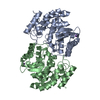
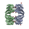
- Structure visualization
| Structure viewer | Molecule: MolmilJmol/JSmol |
|---|
- Downloads & links
-Download
| PDBx/mmCIF format | 5t26.cif.gz | 138.1 KB | Display | PDBx/mmCIF format |
|---|---|---|---|---|
| PDB format | pdb5t26.ent.gz | 104.6 KB | Display | PDB format |
| PDBx/mmJSON format | 5t26.json.gz | Tree view | PDBx/mmJSON format | |
| Others |  Other downloads Other downloads |
-Validation report
| Arichive directory | https://data.pdbj.org/pub/pdb/validation_reports/t2/5t26ftp://data.pdbj.org/pub/pdb/validation_reports/t2/5t26 | HTTPS FTP |
|---|
-Related structure data
| Related structure data |  5t25C  1yxcS C: citing same article ( S: Starting model for refinement |
|---|---|
| Similar structure data |


-Links
 PDBj
PDBj
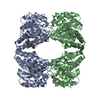
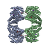
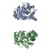
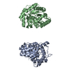
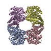
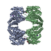
- Assembly
| Deposited unit | 
| |||||||||
|---|---|---|---|---|---|---|---|---|---|---|
| 1 | 
| |||||||||
| Unit cell |
| |||||||||
| Components on special symmetry positions |
|
-Components
| #1: Protein | Mass: 31692.305 Da / Num. of mol.: 2 Source method: isolated from a genetically manipulated source Source: (gene. exp.) Strain: IAI1 / Gene: dapA, ECIAI1_2529 / Production host: References: UniProt: B7M7I1, UniProt: P0A6L2*PLUS, 4-hydroxy-tetrahydrodipicolinate synthase #2: Chemical | ChemComp-NA /   Mass: 22.990 Da / Num. of mol.: 7 / Source method: obtained synthetically / Formula: Na Mass: 22.990 Da / Num. of mol.: 7 / Source method: obtained synthetically / Formula: Na#3: Chemical | ChemComp-TLA / |   Mass: 150.087 Da / Num. of mol.: 1 / Source method: obtained synthetically / Formula: C4H6O6 Mass: 150.087 Da / Num. of mol.: 1 / Source method: obtained synthetically / Formula: C4H6O6#4: Chemical | ChemComp-GOL / |   Mass: 92.094 Da / Num. of mol.: 1 / Source method: obtained synthetically / Formula: C3H8O3 Mass: 92.094 Da / Num. of mol.: 1 / Source method: obtained synthetically / Formula: C3H8O3#5: Water | ChemComp-HOH / |  Mass: 18.015 Da / Num. of mol.: 514 / Source method: isolated from a natural source / Formula: H2O Mass: 18.015 Da / Num. of mol.: 514 / Source method: isolated from a natural source / Formula: H2OHas ligand of interest | Y | |
|---|
-Experimental details
-Experiment
| Experiment | Method: X-RAY DIFFRACTION / Number of used crystals: 1 |
|---|
- Sample preparation
| Crystal | Density Matthews: 2.49 Å3/Da / Density % sol: 50.57 % |
|---|---|
| Crystal grow | Temperature: 293 K / Method: vapor diffusion, hanging drop / pH: 7.1 Details: 20% PEG 3350 200 mM Sodium Tartrate 14.7 mM HEPES pH 7.1 |
-Data collection
| Diffraction | Mean temperature: 100 K / Serial crystal experiment: N |
|---|---|
| Diffraction source | Source: ROTATING ANODE / Type: RIGAKU MICROMAX-007 HF / Wavelength: 1.54178 Å |
| Detector | Type: DECTRIS PILATUS 200K / Detector: PIXEL / Date: Aug 15, 2015 / Details: VeriMaxHF |
| Radiation | Monochromator: multilayer optics / Protocol: SINGLE WAVELENGTH / Monochromatic (M) / Laue (L): M / Scattering type: x-ray |
| Radiation wavelength | Wavelength: 1.54178 Å / Relative weight: 1 |
| Reflection | Resolution: 2.1→81 Å / Num. obs: 35248 / % possible obs: 99.9 % / Redundancy: 6.8 % / Rpim(I) all: 0.052 / Rrim(I) all: 0.142 / Net I/σ(I): 14.4 |
| Reflection shell | Resolution: 2.1→2.14 Å / Num. unique obs: 1813 / CC1/2: 0.968 / Rpim(I) all: 0.236 / Rrim(I) all: 0.462 |
- Processing
| Software |
| ||||||||||||||||||||||||||||||||||||||||||||||||||||||||||||
|---|---|---|---|---|---|---|---|---|---|---|---|---|---|---|---|---|---|---|---|---|---|---|---|---|---|---|---|---|---|---|---|---|---|---|---|---|---|---|---|---|---|---|---|---|---|---|---|---|---|---|---|---|---|---|---|---|---|---|---|---|---|
| Refinement | Method to determine structure: MOLECULAR REPLACEMENT Starting model: 1yxc Resolution: 2.1→27.51 Å / Cor.coef. Fo:Fc: 0.966 / Cor.coef. Fo:Fc free: 0.948 / SU B: 4.37 / SU ML: 0.113 / Cross valid method: THROUGHOUT / σ(F): 0 / ESU R: 0.205 / ESU R Free: 0.166 / Stereochemistry target values: MAXIMUM LIKELIHOOD Details: HYDROGENS HAVE BEEN ADDED IN THE RIDING POSITIONS U VALUES : REFINED INDIVIDUALLY
| ||||||||||||||||||||||||||||||||||||||||||||||||||||||||||||
| Solvent computation | Ion probe radii: 0.8 Å / Shrinkage radii: 0.8 Å / VDW probe radii: 1.2 Å / Solvent model: MASK | ||||||||||||||||||||||||||||||||||||||||||||||||||||||||||||
| Displacement parameters | Biso max: 74.99 Å2 / Biso mean: 16.603 Å2 / Biso min: 6.63 Å2
| ||||||||||||||||||||||||||||||||||||||||||||||||||||||||||||
| Refinement step | Cycle: final / Resolution: 2.1→27.51 Å
| ||||||||||||||||||||||||||||||||||||||||||||||||||||||||||||
| Refine LS restraints |
| ||||||||||||||||||||||||||||||||||||||||||||||||||||||||||||
| LS refinement shell | Resolution: 2.102→2.156 Å / Rfactor Rfree error: 0 / Total num. of bins used: 20
|
