+ データを開く
- 基本情報
基本情報
| 登録情報 | データベース: PDB / ID: 7miz | ||||||
|---|---|---|---|---|---|---|---|
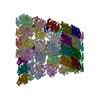
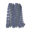
| タイトル | Atomic structure of cortical microtubule from Toxoplasma gondii | ||||||
 要素 要素 |
| ||||||
 キーワード キーワード | STRUCTURAL PROTEIN / cortical / parasite | ||||||
| 機能・相同性 |  機能・相同性情報 機能・相同性情報thioredoxin-disulfide reductase (NADPH) activity / negative regulation of Wnt signaling pathway / negative regulation of protein ubiquitination / structural constituent of cytoskeleton / microtubule cytoskeleton organization / mitotic cell cycle / microtubule binding / 加水分解酵素; 酸無水物に作用; GTPに作用・細胞または細胞小器官の運動に関与 / microtubule / cytoskeleton ...thioredoxin-disulfide reductase (NADPH) activity / negative regulation of Wnt signaling pathway / negative regulation of protein ubiquitination / structural constituent of cytoskeleton / microtubule cytoskeleton organization / mitotic cell cycle / microtubule binding / 加水分解酵素; 酸無水物に作用; GTPに作用・細胞または細胞小器官の運動に関与 / microtubule / cytoskeleton / hydrolase activity / GTPase activity / GTP binding / nucleus / metal ion binding / cytoplasm 類似検索 - 分子機能 | ||||||
| 生物種 |  | ||||||
| 手法 | 電子顕微鏡法 / 単粒子再構成法 / クライオ電子顕微鏡法 / 解像度: 3.4 Å | ||||||
 データ登録者 データ登録者 | Wang, X. / Brown, A. / Sibley, L.D. / Zhang, R. | ||||||
| 資金援助 |  米国, 1件 米国, 1件
| ||||||
 引用 引用 |  ジャーナル: Nat Commun / 年: 2021 ジャーナル: Nat Commun / 年: 2021タイトル: Cryo-EM structure of cortical microtubules from human parasite Toxoplasma gondii identifies their microtubule inner proteins. 著者: Xiangli Wang / Yong Fu / Wandy L Beatty / Meisheng Ma / Alan Brown / L David Sibley / Rui Zhang /  要旨: In living cells, microtubules (MTs) play pleiotropic roles, which require very different mechanical properties. Unlike the dynamic MTs found in the cytoplasm of metazoan cells, the specialized ...In living cells, microtubules (MTs) play pleiotropic roles, which require very different mechanical properties. Unlike the dynamic MTs found in the cytoplasm of metazoan cells, the specialized cortical MTs from Toxoplasma gondii, a prevalent human pathogen, are extraordinarily stable and resistant to detergent and cold treatments. Using single-particle cryo-EM, we determine their ex vivo structure and identify three proteins (TrxL1, TrxL2 and SPM1) as bona fide microtubule inner proteins (MIPs). These three MIPs form a mesh on the luminal surface and simultaneously stabilize the tubulin lattice in both longitudinal and lateral directions. Consistent with previous observations, deletion of the identified MIPs compromises MT stability and integrity under challenges by chemical treatments. We also visualize a small molecule like density at the Taxol-binding site of β-tubulin. Our results provide the structural basis to understand the stability of cortical MTs and suggest an evolutionarily conserved mechanism of MT stabilization from the inside. | ||||||
| 履歴 |
|
- 構造の表示
構造の表示
| ムービー |
 ムービービューア ムービービューア |
|---|---|
| 構造ビューア | 分子:  Molmil Molmil Jmol/JSmol Jmol/JSmol |
- ダウンロードとリンク
- ダウンロード
| PDBx/mmCIF形式 |  7miz.cif.gz 7miz.cif.gz | 5.1 MB | 表示 |  PDBx/mmCIF形式 PDBx/mmCIF形式 |
|---|---|---|---|---|
| PDB形式 |  pdb7miz.ent.gz pdb7miz.ent.gz | 表示 |  PDB形式 PDB形式 | |
| PDBx/mmJSON形式 |  7miz.json.gz 7miz.json.gz | ツリー表示 |  PDBx/mmJSON形式 PDBx/mmJSON形式 | |
| その他 |  その他のダウンロード その他のダウンロード |
-検証レポート
| 文書・要旨 |  7miz_validation.pdf.gz 7miz_validation.pdf.gz | 4.5 MB | 表示 |  wwPDB検証レポート wwPDB検証レポート |
|---|---|---|---|---|
| 文書・詳細版 |  7miz_full_validation.pdf.gz 7miz_full_validation.pdf.gz | 4.7 MB | 表示 | |
| XML形式データ |  7miz_validation.xml.gz 7miz_validation.xml.gz | 644.7 KB | 表示 | |
| CIF形式データ |  7miz_validation.cif.gz 7miz_validation.cif.gz | 985.3 KB | 表示 | |
| アーカイブディレクトリ |  https://data.pdbj.org/pub/pdb/validation_reports/mi/7miz https://data.pdbj.org/pub/pdb/validation_reports/mi/7miz ftp://data.pdbj.org/pub/pdb/validation_reports/mi/7miz ftp://data.pdbj.org/pub/pdb/validation_reports/mi/7miz | HTTPS FTP |
-関連構造データ
- リンク
リンク
- 集合体
集合体
| 登録構造単位 | 
|
|---|---|
| 1 |
|
- 要素
要素
-タンパク質 , 5種, 100分子 01234567891011121314151617181920212223A0A2A4A6A8B0...
| #1: タンパク質 | 分子量: 38845.750 Da / 分子数: 24 / 由来タイプ: 天然 / 由来: (天然)  #2: タンパク質 | 分子量: 50166.645 Da / 分子数: 26 / 由来タイプ: 天然 / 由来: (天然)  #3: タンパク質 | 分子量: 50119.121 Da / 分子数: 26 / 由来タイプ: 天然 / 由来: (天然)  #4: タンパク質 | 分子量: 24939.332 Da / 分子数: 20 / 由来タイプ: 天然 / 由来: (天然)  #5: タンパク質 | 分子量: 21687.934 Da / 分子数: 4 / 由来タイプ: 天然 / 由来: (天然)  |
|---|
-非ポリマー , 3種, 78分子 


| #6: 化合物 | ChemComp-GTP / #7: 化合物 | ChemComp-MG / #8: 化合物 | ChemComp-GDP / |
|---|
-詳細
| 研究の焦点であるリガンドがあるか | N |
|---|---|
| Has protein modification | Y |
-実験情報
-実験
| 実験 | 手法: 電子顕微鏡法 |
|---|---|
| EM実験 | 試料の集合状態: FILAMENT / 3次元再構成法: 単粒子再構成法 |
- 試料調製
試料調製
| 構成要素 | 名称: cortical microtubule with associated MIPs / タイプ: COMPLEX / Entity ID: #1-#5 / 由来: NATURAL |
|---|---|
| 分子量 | 実験値: NO |
| 由来(天然) | 生物種:  |
| 緩衝液 | pH: 7.4 |
| 試料 | 包埋: NO / シャドウイング: NO / 染色: NO / 凍結: YES |
| 試料支持 | グリッドの材料: COPPER / グリッドのサイズ: 300 divisions/in. / グリッドのタイプ: Quantifoil R2/2 |
| 急速凍結 | 装置: FEI VITROBOT MARK IV / 凍結剤: ETHANE / 湿度: 95 % / 凍結前の試料温度: 289 K / 詳細: blot for 4 seconds with a blot force of -15 |
- 電子顕微鏡撮影
| 実験機器 |  モデル: Titan Krios / 画像提供: FEI Company |
|---|---|
| 顕微鏡 | モデル: FEI TITAN KRIOS |
| 電子銃 | 電子線源:  FIELD EMISSION GUN / 加速電圧: 300 kV / 照射モード: FLOOD BEAM FIELD EMISSION GUN / 加速電圧: 300 kV / 照射モード: FLOOD BEAM |
| 電子レンズ | モード: BRIGHT FIELD / 倍率(公称値): 105000 X / 最大 デフォーカス(公称値): 3500 nm / Calibrated defocus min: 1000 nm / Cs: 0.01 mm / アライメント法: ZEMLIN TABLEAU |
| 試料ホルダ | 凍結剤: NITROGEN 試料ホルダーモデル: FEI TITAN KRIOS AUTOGRID HOLDER |
| 撮影 | 平均露光時間: 9 sec. / 電子線照射量: 63.7 e/Å2 フィルム・検出器のモデル: GATAN K3 BIOQUANTUM (6k x 4k) 撮影したグリッド数: 2 / 実像数: 9231 詳細: Images were collected in counting mode, with an exposure rate of 8.5 electrons/pixel/s on the detector camera. |
| 画像スキャン | 横: 3838 / 縦: 3710 |
- 解析
解析
| EMソフトウェア |
| ||||||||||||||||||||||||||||||||||||||||||||||||||||
|---|---|---|---|---|---|---|---|---|---|---|---|---|---|---|---|---|---|---|---|---|---|---|---|---|---|---|---|---|---|---|---|---|---|---|---|---|---|---|---|---|---|---|---|---|---|---|---|---|---|---|---|---|---|
| CTF補正 | 詳細: CTF amplitude correction was performed as part of the 3D reconstruction. タイプ: PHASE FLIPPING AND AMPLITUDE CORRECTION | ||||||||||||||||||||||||||||||||||||||||||||||||||||
| 粒子像の選択 | 選択した粒子像数: 536132 詳細: Cortical MTs were manually picked from the motion-corrected micrographs using the APPION image-processing suite. The selected MT segments were computationally cut into overlapping boxes ...詳細: Cortical MTs were manually picked from the motion-corrected micrographs using the APPION image-processing suite. The selected MT segments were computationally cut into overlapping boxes (512x512 pixels) with an 8-nm non-overlapping region (step size) between adjacent boxes (corresponding to the length of a tubulin heterodimer). We refer to these boxed images as 8-nm MT particles. | ||||||||||||||||||||||||||||||||||||||||||||||||||||
| 対称性 | 点対称性: C1 (非対称) | ||||||||||||||||||||||||||||||||||||||||||||||||||||
| 3次元再構成 | 解像度: 3.4 Å / 解像度の算出法: FSC 0.143 CUT-OFF / 粒子像の数: 220139 / アルゴリズム: FOURIER SPACE / 対称性のタイプ: POINT | ||||||||||||||||||||||||||||||||||||||||||||||||||||
| 原子モデル構築 | プロトコル: AB INITIO MODEL 詳細: Secondary structure, Ramachandran, and rotamer restraints were applied during refinement. The nonbonded_weight value was set to 500 to reduce the clashes between atoms. | ||||||||||||||||||||||||||||||||||||||||||||||||||||
| 原子モデル構築 |
|
 ムービー
ムービー コントローラー


 PDBj
PDBj


