 Movie
Movie Controller


+ Open data

- Basic information
| Entry | Database: PDB / ID: 7mgy | ||||||
|---|---|---|---|---|---|---|---|
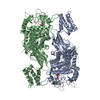
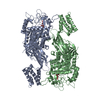
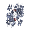
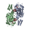
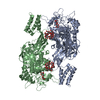
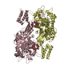
| Title | Sco GlgEI-V279S in complex with 4-alpha-glucoside of valienamine | ||||||
 Components Components | Alpha-1,4-glucan:maltose-1-phosphate maltosyltransferase 1 | ||||||
 Keywords Keywords | TRANSFERASE / CAZy glycoside hydrolase (GH)-like phosphorylase GH13_3 family / elongation of cytosolic glucan chains / maltosyl transferase | ||||||
| Function / homology |  Function and homology information Function and homology informationstarch synthase (maltosyl-transferring) / alpha-glucan biosynthetic process / hexosyltransferase activity / alpha-amylase activity / oligosaccharide catabolic process Similarity search - Function | ||||||
| Biological species |  Streptomyces coelicolor (bacteria) Streptomyces coelicolor (bacteria) | ||||||
| Method |  X-RAY DIFFRACTION / SYNCHROTRON / MOLECULAR REPLACEMENT / Resolution: 1.83 Å X-RAY DIFFRACTION / SYNCHROTRON / MOLECULAR REPLACEMENT / Resolution: 1.83 Å | ||||||
 Authors Authors | Jayasinghe, T.D. / Ronning, D.R. | ||||||
| Funding support |  United States, 1items United States, 1items
| ||||||
 Citation Citation | Journal: Sci Rep / Year: 2021 Title: Stereoselective synthesis of a 4-⍺-glucoside of valienamine and its X-ray structure in complex with Streptomyces coelicolor GlgE1-V279S. Authors: Si, A. / Jayasinghe, T.D. / Thanvi, R. / Ronning, D.R. / Sucheck, S.J. | ||||||
| History |
|
- Structure visualization
| Structure viewer | Molecule: MolmilJmol/JSmol |
|---|
- Downloads & links
-Download
| PDBx/mmCIF format | 7mgy.cif.gz | 300 KB | Display | PDBx/mmCIF format |
|---|---|---|---|---|
| PDB format | pdb7mgy.ent.gz | 238 KB | Display | PDB format |
| PDBx/mmJSON format | 7mgy.json.gz | Tree view | PDBx/mmJSON format | |
| Others |  Other downloads Other downloads |
-Validation report
| Arichive directory | https://data.pdbj.org/pub/pdb/validation_reports/mg/7mgyftp://data.pdbj.org/pub/pdb/validation_reports/mg/7mgy | HTTPS FTP |
|---|
-Related structure data
| Related structure data | 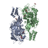 7melC 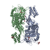 4u31S S: Starting model for refinement C: citing same article ( |
|---|---|
| Similar structure data |


-Links
 PDBj
PDBj

- Assembly
| Deposited unit | 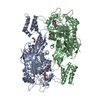
| ||||||||
|---|---|---|---|---|---|---|---|---|---|
| 1 |
| ||||||||
| Unit cell |
|
-Components
| #1: Protein | Mass: 76394.547 Da / Num. of mol.: 2 Source method: isolated from a genetically manipulated source Source: (gene. exp.) Streptomyces coelicolor (bacteria) / Strain: ATCC BAA-471 / A3(2) / M145 / Gene: glgE1, pep1, pep1A, pep1I, SCO5443, SC6A11.19c / Production host: References: UniProt: Q9L1K2, starch synthase (maltosyl-transferring) #2: Chemical |   Mass: 337.323 Da / Num. of mol.: 2 / Source method: obtained synthetically / Formula: C13H23NO9 / Feature type: SUBJECT OF INVESTIGATION Mass: 337.323 Da / Num. of mol.: 2 / Source method: obtained synthetically / Formula: C13H23NO9 / Feature type: SUBJECT OF INVESTIGATION#3: Chemical | ChemComp-PGE /   Mass: 150.173 Da / Num. of mol.: 4 / Source method: obtained synthetically / Formula: C6H14O4 Mass: 150.173 Da / Num. of mol.: 4 / Source method: obtained synthetically / Formula: C6H14O4#4: Chemical | ChemComp-PEG / |   Mass: 106.120 Da / Num. of mol.: 1 / Source method: obtained synthetically / Formula: C4H10O3 Mass: 106.120 Da / Num. of mol.: 1 / Source method: obtained synthetically / Formula: C4H10O3#5: Water | ChemComp-HOH / |  Mass: 18.015 Da / Num. of mol.: 1404 / Source method: isolated from a natural source / Formula: H2O Mass: 18.015 Da / Num. of mol.: 1404 / Source method: isolated from a natural source / Formula: H2OHas ligand of interest | Y | |
|---|
-Experimental details
-Experiment
| Experiment | Method: X-RAY DIFFRACTION / Number of used crystals: 1 |
|---|
- Sample preparation
| Crystal | Density Matthews: 3.23 Å3/Da / Density % sol: 61.9 % |
|---|---|
| Crystal grow | Temperature: 298.15 K / Method: vapor diffusion, hanging drop / pH: 7 / Details: 0.2 mM Sodium citrate pH 7 and 10 % PEG 3350 |
-Data collection
| Diffraction | Mean temperature: 100 K / Serial crystal experiment: N |
|---|---|
| Diffraction source | Source: SYNCHROTRON / Site: APS / Beamline: 21-ID-F / Wavelength: 0.9787 Å |
| Detector | Type: MARMOSAIC 225 mm CCD / Detector: CCD / Date: Jul 31, 2020 |
| Radiation | Protocol: SINGLE WAVELENGTH / Monochromatic (M) / Laue (L): M / Scattering type: x-ray |
| Radiation wavelength | Wavelength: 0.9787 Å / Relative weight: 1 |
| Reflection | Resolution: 1.83→55.48 Å / Num. obs: 176156 / % possible obs: 93.14 % / Redundancy: 16.3 % / Biso Wilson estimate: 31.12 Å2 / CC1/2: 0.998 / Rmerge(I) obs: 0.1802 / Rpim(I) all: 0.04559 / Rrim(I) all: 0.186 / Net I/σ(I): 9.28 |
| Reflection shell | Resolution: 1.83→1.896 Å / Rmerge(I) obs: 6.231 / Mean I/σ(I) obs: 0.21 / Num. unique obs: 17390 / CC1/2: 0.436 / Rrim(I) all: 6.499 |
- Processing
| Software |
| ||||||||||||||||||||||||||||||||||||||||||||||||||||||||||||||||||||||||||||||||||||||||||||||||||
|---|---|---|---|---|---|---|---|---|---|---|---|---|---|---|---|---|---|---|---|---|---|---|---|---|---|---|---|---|---|---|---|---|---|---|---|---|---|---|---|---|---|---|---|---|---|---|---|---|---|---|---|---|---|---|---|---|---|---|---|---|---|---|---|---|---|---|---|---|---|---|---|---|---|---|---|---|---|---|---|---|---|---|---|---|---|---|---|---|---|---|---|---|---|---|---|---|---|---|---|
| Refinement | Method to determine structure: MOLECULAR REPLACEMENT Starting model: 4U31 Resolution: 1.83→55.48 Å / SU ML: 0.23 / Cross valid method: THROUGHOUT / σ(F): 1.33 / Phase error: 22.65 / Stereochemistry target values: ML
| ||||||||||||||||||||||||||||||||||||||||||||||||||||||||||||||||||||||||||||||||||||||||||||||||||
| Solvent computation | Shrinkage radii: 0.9 Å / VDW probe radii: 1.11 Å / Solvent model: FLAT BULK SOLVENT MODEL | ||||||||||||||||||||||||||||||||||||||||||||||||||||||||||||||||||||||||||||||||||||||||||||||||||
| Displacement parameters | Biso max: 93.83 Å2 / Biso mean: 33.6943 Å2 / Biso min: 17.66 Å2 | ||||||||||||||||||||||||||||||||||||||||||||||||||||||||||||||||||||||||||||||||||||||||||||||||||
| Refinement step | Cycle: final / Resolution: 1.83→55.48 Å
| ||||||||||||||||||||||||||||||||||||||||||||||||||||||||||||||||||||||||||||||||||||||||||||||||||
| LS refinement shell | Refine-ID: X-RAY DIFFRACTION / Rfactor Rfree error: 0 / Total num. of bins used: 13
|
