 Movie
Movie Controller

[English] 日本語
 Yorodumi
- PDB-5vsj: Sco GlgEI-V279S in complex with a pyrolidene-based ethyl-phosphon... -
+ Open data

- Basic information
| Entry | Database: PDB / ID: 5vsj | ||||||
|---|---|---|---|---|---|---|---|
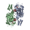
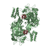
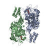
| Title | Sco GlgEI-V279S in complex with a pyrolidene-based ethyl-phosphonate compound | ||||||
 Components Components | Alpha-1,4-glucan:maltose-1-phosphate maltosyltransferase 1 | ||||||
 Keywords Keywords | TRANSFERASE / Streptomyces coelicolor / Maltosyltransferase / transition state analogue / Alpha-1 / 4-glucan | ||||||
| Function / homology |  Function and homology information Function and homology informationstarch synthase (maltosyl-transferring) / alpha-glucan biosynthetic process / hexosyltransferase activity / alpha-amylase activity / oligosaccharide catabolic process Similarity search - Function | ||||||
| Biological species |  Streptomyces coelicolor (bacteria) Streptomyces coelicolor (bacteria) | ||||||
| Method |  X-RAY DIFFRACTION / SYNCHROTRON / MOLECULAR REPLACEMENT / Resolution: 2.456 Å X-RAY DIFFRACTION / SYNCHROTRON / MOLECULAR REPLACEMENT / Resolution: 2.456 Å | ||||||
 Authors Authors | Petit, C. / Ronning, D.R. | ||||||
| Funding support |  United States, 1items United States, 1items
| ||||||
 Citation Citation | Journal: Org. Biomol. Chem. / Year: 2017 Title: Zwitterionic pyrrolidene-phosphonates: inhibitors of the glycoside hydrolase-like phosphorylase Streptomyces coelicolor GlgEI-V279S. Authors: Veleti, S.K. / Petit, C. / Ronning, D.R. / Sucheck, S.J. #1: Journal: Org. Biomol. Chem. / Year: 2017Title: Zwitterionic pyrrolidene-phosphonates: inhibitors of the glycoside hydrolase-like phosphorylase Streptomyces coelicolor GlgEI-V279S. Authors: Veleti, S.K. / Petit, C. / Ronning, D.R. / Sucheck, S.J. | ||||||
| History |
|
- Structure visualization
| Structure viewer | Molecule: MolmilJmol/JSmol |
|---|
- Downloads & links
-Download
| PDBx/mmCIF format | 5vsj.cif.gz | 272.4 KB | Display | PDBx/mmCIF format |
|---|---|---|---|---|
| PDB format | pdb5vsj.ent.gz | 218.6 KB | Display | PDB format |
| PDBx/mmJSON format | 5vsj.json.gz | Tree view | PDBx/mmJSON format | |
| Others |  Other downloads Other downloads |
-Validation report
| Arichive directory | https://data.pdbj.org/pub/pdb/validation_reports/vs/5vsjftp://data.pdbj.org/pub/pdb/validation_reports/vs/5vsj | HTTPS FTP |
|---|
-Related structure data
| Related structure data |  5vt4C  4cn1S C: citing same article ( S: Starting model for refinement |
|---|---|
| Similar structure data |


-Links
 PDBj
PDBj

- Assembly
| Deposited unit | 
| ||||||||
|---|---|---|---|---|---|---|---|---|---|
| 1 |
| ||||||||
| Unit cell |
|
-Components
| #1: Protein | Mass: 74179.133 Da / Num. of mol.: 2 / Mutation: V279S Source method: isolated from a genetically manipulated source Source: (gene. exp.) Streptomyces coelicolor (strain ATCC BAA-471 / A3(2) / M145) (bacteria)Strain: ATCC BAA-471 / A3(2) / M145 / Gene: glgE1, pep1, pep1A, pep1I, SCO5443, SC6A11.19c / Cell line (production host): BL21 / Production host: References: UniProt: Q9L1K2, starch synthase (maltosyl-transferring) #2: Chemical |   Mass: 433.345 Da / Num. of mol.: 2 / Source method: obtained synthetically / Formula: C14H28NO12P Mass: 433.345 Da / Num. of mol.: 2 / Source method: obtained synthetically / Formula: C14H28NO12P#3: Chemical | ChemComp-TRS / |   Mass: 122.143 Da / Num. of mol.: 1 / Source method: isolated from a natural source / Formula: C4H12NO3 / Comment: pH buffer*YM Mass: 122.143 Da / Num. of mol.: 1 / Source method: isolated from a natural source / Formula: C4H12NO3 / Comment: pH buffer*YM#4: Water | ChemComp-HOH / |  Mass: 18.015 Da / Num. of mol.: 359 / Source method: isolated from a natural source / Formula: H2O Mass: 18.015 Da / Num. of mol.: 359 / Source method: isolated from a natural source / Formula: H2O |
|---|
-Experimental details
-Experiment
| Experiment | Method: X-RAY DIFFRACTION / Number of used crystals: 1 |
|---|
- Sample preparation
| Crystal | Density Matthews: 3.47 Å3/Da / Density % sol: 64.51 % / Description: Diamond |
|---|---|
| Crystal grow | Temperature: 293 K / Method: vapor diffusion, hanging drop / pH: 7 / Details: 0.2 M sodium citrate pH 7.0 and 10 % PEG 3,350 |
-Data collection
| Diffraction | Mean temperature: 100 K |
|---|---|
| Diffraction source | Source: SYNCHROTRON / Site: APS / Beamline: 21-ID-F / Wavelength: 0.97872 Å |
| Detector | Type: MARMOSAIC 225 mm CCD / Detector: CCD / Date: Jul 24, 2015 |
| Radiation | Protocol: SINGLE WAVELENGTH / Monochromatic (M) / Laue (L): M / Scattering type: x-ray |
| Radiation wavelength | Wavelength: 0.97872 Å / Relative weight: 1 |
| Reflection | Resolution: 2.45→45.878 Å / Num. obs: 76413 / % possible obs: 99.1 % / Redundancy: 12.7 % / Net I/σ(I): 14.84 |
| Reflection shell | Redundancy: 13.3 % / Num. unique obs: 7224 / % possible all: 95.1 |
- Processing
| Software |
| |||||||||||||||||||||||||||||||||||||||||||||||||||||||||||||||||||||||||||||||||||||||||||||||||||||||||
|---|---|---|---|---|---|---|---|---|---|---|---|---|---|---|---|---|---|---|---|---|---|---|---|---|---|---|---|---|---|---|---|---|---|---|---|---|---|---|---|---|---|---|---|---|---|---|---|---|---|---|---|---|---|---|---|---|---|---|---|---|---|---|---|---|---|---|---|---|---|---|---|---|---|---|---|---|---|---|---|---|---|---|---|---|---|---|---|---|---|---|---|---|---|---|---|---|---|---|---|---|---|---|---|---|---|---|
| Refinement | Method to determine structure: MOLECULAR REPLACEMENT Starting model: 4CN1 Resolution: 2.456→45.878 Å / SU ML: 0.32 / Cross valid method: NONE / σ(F): 1.34 / Phase error: 25.09 / Stereochemistry target values: ML
| |||||||||||||||||||||||||||||||||||||||||||||||||||||||||||||||||||||||||||||||||||||||||||||||||||||||||
| Solvent computation | Shrinkage radii: 0.9 Å / VDW probe radii: 1.11 Å / Solvent model: FLAT BULK SOLVENT MODEL | |||||||||||||||||||||||||||||||||||||||||||||||||||||||||||||||||||||||||||||||||||||||||||||||||||||||||
| Refinement step | Cycle: LAST / Resolution: 2.456→45.878 Å
| |||||||||||||||||||||||||||||||||||||||||||||||||||||||||||||||||||||||||||||||||||||||||||||||||||||||||
| Refine LS restraints |
| |||||||||||||||||||||||||||||||||||||||||||||||||||||||||||||||||||||||||||||||||||||||||||||||||||||||||
| LS refinement shell |
|
