 Movie
Movie Controller


+ Open data

- Basic information
| Entry | Database: PDB / ID: 7mbk | ||||||
|---|---|---|---|---|---|---|---|
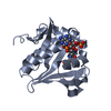
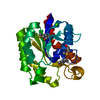
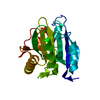
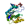
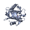
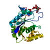
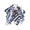
| Title | N-terminal domain of mouse surfactant protein B, 6W mutant | ||||||
 Components Components | Pulmonary surfactant-associated protein B | ||||||
 Keywords Keywords | SURFACTANT PROTEIN | ||||||
| Function / homology |  Function and homology information Function and homology informationSurfactant metabolism / alveolar lamellar body / sphingolipid metabolic process / respiratory gaseous exchange by respiratory system / multivesicular body / extracellular matrix / lysosome / : / membrane / cytoplasm Similarity search - Function | ||||||
| Biological species |  | ||||||
| Method |  X-RAY DIFFRACTION / SYNCHROTRON / MOLECULAR REPLACEMENT / Resolution: 2.17 Å X-RAY DIFFRACTION / SYNCHROTRON / MOLECULAR REPLACEMENT / Resolution: 2.17 Å | ||||||
 Authors Authors | Milicic, G. / Rapoport, T.A. | ||||||
| Funding support |  United States, 1items United States, 1items
| ||||||
 Citation Citation | Journal: Mol.Cell / Year: 2021 Title: Mechanism of Lamellar Body Formation by Lung Surfactant Protein B. Authors: Sever, N. / Milicic, G. / Bodnar, N.O. / Wu, X. / Rapoport, T.A. | ||||||
| History |
|
- Structure visualization
| Structure viewer | Molecule: MolmilJmol/JSmol |
|---|
- Downloads & links
-Download
| PDBx/mmCIF format | 7mbk.cif.gz | 80.6 KB | Display | PDBx/mmCIF format |
|---|---|---|---|---|
| PDB format | pdb7mbk.ent.gz | 56.1 KB | Display | PDB format |
| PDBx/mmJSON format | 7mbk.json.gz | Tree view | PDBx/mmJSON format | |
| Others |  Other downloads Other downloads |
-Validation report
| Arichive directory | https://data.pdbj.org/pub/pdb/validation_reports/mb/7mbkftp://data.pdbj.org/pub/pdb/validation_reports/mb/7mbk | HTTPS FTP |
|---|
-Related structure data
| Related structure data | 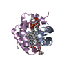 6vynSC 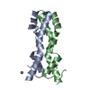 6vz0C  6vzdC 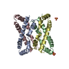 6vzeC 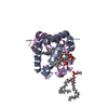 6w1bC S: Starting model for refinement C: citing same article ( |
|---|---|
| Similar structure data |


-Links
 PDBj
PDBj- Assembly
| Deposited unit | 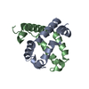
| ||||||||||||
|---|---|---|---|---|---|---|---|---|---|---|---|---|---|
| 1 |
| ||||||||||||
| Unit cell |
|
-Components
| #1: Protein | Mass: 10429.968 Da / Num. of mol.: 2 / Fragment: N-terminal domain, UNP residues 61-146 / Mutation: L35W, L44W, V48W, V62W, I75W, V79W Source method: isolated from a genetically manipulated source Source: (gene. exp.)  #2: Water | ChemComp-HOH / |  Mass: 18.015 Da / Num. of mol.: 12 / Source method: isolated from a natural source / Formula: H2O Mass: 18.015 Da / Num. of mol.: 12 / Source method: isolated from a natural source / Formula: H2OHas protein modification | Y | |
|---|
-Experimental details
-Experiment
| Experiment | Method: X-RAY DIFFRACTION / Number of used crystals: 1 |
|---|
- Sample preparation
| Crystal | Density Matthews: 2.16 Å3/Da / Density % sol: 42.99 % |
|---|---|
| Crystal grow | Temperature: 293.15 K / Method: vapor diffusion, hanging drop / pH: 5.5 Details: 0.1 M ammonium sulfate, 0.05 M magnesium sulfate, 0.1 M sodium citrate, 16.5 % PEG smear medium |
-Data collection
| Diffraction | Mean temperature: 100 K / Serial crystal experiment: N |
|---|---|
| Diffraction source | Source: SYNCHROTRON / Site: APS / Beamline: 24-ID-C / Wavelength: 1.2827 Å |
| Detector | Type: DECTRIS PILATUS 6M-F / Detector: PIXEL / Date: Jun 13, 2019 |
| Radiation | Protocol: SINGLE WAVELENGTH / Monochromatic (M) / Laue (L): M / Scattering type: x-ray |
| Radiation wavelength | Wavelength: 1.2827 Å / Relative weight: 1 |
| Reflection | Resolution: 2.17→47.45 Å / Num. obs: 9974 / % possible obs: 96.6 % / Redundancy: 20 % / Biso Wilson estimate: 50.16 Å2 / Rrim(I) all: 0.147 / Net I/σ(I): 23.1 |
| Reflection shell | Resolution: 2.17→2.29 Å / Num. unique obs: 375 / Rrim(I) all: 0.148 |
- Processing
| Software |
| ||||||||||||||||||||||||||||||||||||||||||||||||||||||||
|---|---|---|---|---|---|---|---|---|---|---|---|---|---|---|---|---|---|---|---|---|---|---|---|---|---|---|---|---|---|---|---|---|---|---|---|---|---|---|---|---|---|---|---|---|---|---|---|---|---|---|---|---|---|---|---|---|---|
| Refinement | Method to determine structure: MOLECULAR REPLACEMENT Starting model: 6vyn Resolution: 2.17→47.45 Å / SU ML: 0.2981 / Cross valid method: FREE R-VALUE / σ(F): 0 / Phase error: 28.3877 Stereochemistry target values: GeoStd + Monomer Library + CDL v1.2
| ||||||||||||||||||||||||||||||||||||||||||||||||||||||||
| Solvent computation | Shrinkage radii: 0.9 Å / VDW probe radii: 1.11 Å / Solvent model: FLAT BULK SOLVENT MODEL | ||||||||||||||||||||||||||||||||||||||||||||||||||||||||
| Displacement parameters | Biso mean: 60.79 Å2 | ||||||||||||||||||||||||||||||||||||||||||||||||||||||||
| Refinement step | Cycle: LAST / Resolution: 2.17→47.45 Å
| ||||||||||||||||||||||||||||||||||||||||||||||||||||||||
| Refine LS restraints |
| ||||||||||||||||||||||||||||||||||||||||||||||||||||||||
| LS refinement shell |
|
