 Movie
Movie Controller

[English] 日本語
 Yorodumi
- PDB-7m4r: Structural basis for SARS-CoV-2 envelope protein in recognition o... -
+ Open data

- Basic information
| Entry | Database: PDB / ID: 7m4r | ||||||||||||||||||||||||||||||||||||
|---|---|---|---|---|---|---|---|---|---|---|---|---|---|---|---|---|---|---|---|---|---|---|---|---|---|---|---|---|---|---|---|---|---|---|---|---|---|
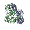
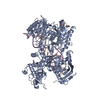
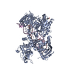
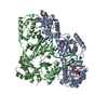
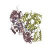
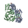
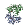
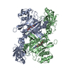
| Title | Structural basis for SARS-CoV-2 envelope protein in recognition of human cell junction protein PALS1 | ||||||||||||||||||||||||||||||||||||
 Components Components |
| ||||||||||||||||||||||||||||||||||||
 Keywords Keywords | CELL ADHESION/VIRAL PROTEIN / SARS-CoV-2 envelope protein / PDZ-binding motif / complex / pathogen-host interaction / CELL ADHESION-VIRAL PROTEIN complex | ||||||||||||||||||||||||||||||||||||
| Function / homology |  Function and homology information Function and homology informationprotein localization to myelin sheath abaxonal region / disruption of cellular anatomical structure in another organism / SARS-CoV-1 targets PDZ proteins in cell-cell junction / symbiont-mediated perturbation of host cell endomembrane system / establishment or maintenance of polarity of embryonic epithelium / viral budding from Golgi membrane / morphogenesis of an epithelial sheet / myelin assembly / Tight junction interactions / regulation of transforming growth factor beta receptor signaling pathway ...protein localization to myelin sheath abaxonal region / disruption of cellular anatomical structure in another organism / SARS-CoV-1 targets PDZ proteins in cell-cell junction / symbiont-mediated perturbation of host cell endomembrane system / establishment or maintenance of polarity of embryonic epithelium / viral budding from Golgi membrane / morphogenesis of an epithelial sheet / myelin assembly / Tight junction interactions / regulation of transforming growth factor beta receptor signaling pathway / SARS-CoV-2 targets PDZ proteins in cell-cell junction / myelin sheath adaxonal region / Regulation of gap junction activity / lateral loop / Lectin pathway of complement activation / establishment or maintenance of epithelial cell apical/basal polarity / peripheral nervous system myelin maintenance / Schmidt-Lanterman incisure / apical junction complex / generation of neurons / central nervous system neuron development / Initial triggering of complement / host cell Golgi membrane / bicellular tight junction / endoplasmic reticulum-Golgi intermediate compartment membrane / Maturation of protein E / protein localization to plasma membrane / adherens junction / cerebral cortex development / cell junction / Translation of Structural Proteins / Virion Assembly and Release / Induction of Cell-Cell Fusion / Attachment and Entry / perikaryon / apical plasma membrane / protein domain specific binding / axon / SARS-CoV-2 activates/modulates innate and adaptive immune responses / Golgi apparatus / virion membrane / protein-containing complex / extracellular exosome / ATP binding / membrane / identical protein binding / plasma membrane / cytoplasm Similarity search - Function | ||||||||||||||||||||||||||||||||||||
| Biological species |  Homo sapiens (human) Homo sapiens (human)  Severe acute respiratory syndrome coronavirus 2 Severe acute respiratory syndrome coronavirus 2 | ||||||||||||||||||||||||||||||||||||
| Method | ELECTRON MICROSCOPY / single particle reconstruction / cryo EM / Resolution: 3.65 Å | ||||||||||||||||||||||||||||||||||||
 Authors Authors | Liu, Q. / Chai, J. | ||||||||||||||||||||||||||||||||||||
 Citation Citation | Journal: Nat Commun / Year: 2021 Title: Structural basis for SARS-CoV-2 envelope protein recognition of human cell junction protein PALS1. Authors: Jin Chai / Yuanheng Cai / Changxu Pang / Liguo Wang / Sean McSweeney / John Shanklin / Qun Liu /  Abstract: The COVID-19 pandemic, caused by the SARS-CoV-2 virus, has created global health and economic emergencies. SARS-CoV-2 viruses promote their own spread and virulence by hijacking human proteins, which ...The COVID-19 pandemic, caused by the SARS-CoV-2 virus, has created global health and economic emergencies. SARS-CoV-2 viruses promote their own spread and virulence by hijacking human proteins, which occurs through viral protein recognition of human targets. To understand the structural basis for SARS-CoV-2 viral-host protein recognition, here we use cryo-electron microscopy (cryo-EM) to determine a complex structure of the human cell junction protein PALS1 and SARS-CoV-2 viral envelope (E) protein. Our reported structure shows that the E protein C-terminal DLLV motif recognizes a pocket formed exclusively by hydrophobic residues from the PDZ and SH3 domains of PALS1. Our structural analysis provides an explanation for the observation that the viral E protein recruits PALS1 from lung epithelial cell junctions. In addition, our structure provides novel targets for peptide- and small-molecule inhibitors that could block the PALS1-E interactions to reduce E-mediated virulence. | ||||||||||||||||||||||||||||||||||||
| History |
|
- Structure visualization
| Movie |
Movie viewer |
|---|---|
| Structure viewer | Molecule: MolmilJmol/JSmol |
- Downloads & links
-Download
| PDBx/mmCIF format | 7m4r.cif.gz | 165 KB | Display | PDBx/mmCIF format |
|---|---|---|---|---|
| PDB format | pdb7m4r.ent.gz | 120.7 KB | Display | PDB format |
| PDBx/mmJSON format | 7m4r.json.gz | Tree view | PDBx/mmJSON format | |
| Others |  Other downloads Other downloads |
-Validation report
| Arichive directory | https://data.pdbj.org/pub/pdb/validation_reports/m4/7m4rftp://data.pdbj.org/pub/pdb/validation_reports/m4/7m4r | HTTPS FTP |
|---|
-Related structure data
| Related structure data |  23665MC M: map data used to model this data C: citing same article ( |
|---|---|
| Similar structure data |


-Links
 PDBj
PDBj


- Assembly
| Deposited unit | 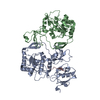
|
|---|---|
| 1 |
|
-Components
| #1: Protein | Mass: 44780.504 Da / Num. of mol.: 2 / Fragment: UNP residues 236-410,461-675 Source method: isolated from a genetically manipulated source Source: (gene. exp.) Homo sapiens (human) / Gene: MPP5, PALS1 / Production host:  #2: Protein/peptide | | Mass: 2062.395 Da / Num. of mol.: 1 / Fragment: UNP residues 58-75 / Source method: obtained synthetically Source: (synth.) Severe acute respiratory syndrome coronavirus 2References: UniProt: P0DTC4 Has protein modification | N | |
|---|
-Experimental details
-Experiment
| Experiment | Method: ELECTRON MICROSCOPY |
|---|---|
| EM experiment | Aggregation state: PARTICLE / 3D reconstruction method: single particle reconstruction |
- Sample preparation
| Component | Name: Complex structure of SARS-CoV-2 envelope protein Ec18 and human PALS1 PSG domains Type: COMPLEX / Entity ID: all / Source: MULTIPLE SOURCES |
|---|---|
| Source (natural) | Organism: Homo sapiens (human) |
| Source (recombinant) | Organism: |
| Buffer solution | pH: 7.5 |
| Specimen | Embedding applied: NO / Shadowing applied: NO / Staining applied: NO / Vitrification applied: YES |
| Vitrification | Cryogen name: ETHANE |
- Electron microscopy imaging
| Experimental equipment |  Model: Titan Krios / Image courtesy: FEI Company |
|---|---|
| Microscopy | Model: FEI TITAN KRIOS |
| Electron gun | Electron source:  FIELD EMISSION GUN / Accelerating voltage: 300 kV / Illumination mode: FLOOD BEAM FIELD EMISSION GUN / Accelerating voltage: 300 kV / Illumination mode: FLOOD BEAM |
| Electron lens | Mode: BRIGHT FIELD |
| Image recording | Electron dose: 64 e/Å2 / Film or detector model: GATAN K3 BIOQUANTUM (6k x 4k) |
- Processing
| Software | Name: PHENIX / Version: 1.18.2_3874: / Classification: refinement | ||||||||||||||||||||||||
|---|---|---|---|---|---|---|---|---|---|---|---|---|---|---|---|---|---|---|---|---|---|---|---|---|---|
| EM software | Name: PHENIX / Category: model refinement | ||||||||||||||||||||||||
| CTF correction | Type: PHASE FLIPPING AND AMPLITUDE CORRECTION | ||||||||||||||||||||||||
| 3D reconstruction | Resolution: 3.65 Å / Resolution method: FSC 0.143 CUT-OFF / Num. of particles: 47615 / Symmetry type: POINT | ||||||||||||||||||||||||
| Refine LS restraints |
|
