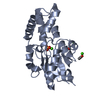
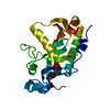
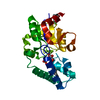
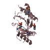
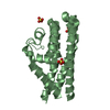
Guanylatecyclaseactivator1A / Guanylyl cyclase-activating protein 1
Mass: 22489.383 Da / Num. of mol.: 1 Source method: isolated from a genetically manipulated source Details: The initial MET at residue 1 gets removed during the myristoylation of the protein. The myristoyl group is covalently attached (N-acyl linkage) to the amino group of the GLY at residue 2. Source: (gene. exp.) Danio rerio (zebrafish) / Gene: guca1e, GUCA1E / Production host: Escherichia coli (E. coli) / References: UniProt: Q5MAC8
Mass: 24.305 Da / Num. of mol.: 1 / Source method: obtained synthetically / Formula: Mg
Has ligand of interest
N
-
Experimental details
-
Experiment
Experiment
Method: SOLUTION NMR
NMR experiment
Conditions-ID
Experiment-ID
Solution-ID
Sample state
Spectrometer-ID
Type
1
1
1
isotropic
1
2D 1H-15N HSQC
2
2
2
isotropic
1
2D 1H-13C HSQC aliphatic
2
3
2
isotropic
1
3D HN(CA)CB
2
4
2
isotropic
1
3DCBCA(CO)NH
2
5
2
isotropic
1
3D (H)CCH-TOCSY
2
6
2
isotropic
1
3D 1H-13C NOESY aliphatic
-
Sample preparation
Details
Type
Solution-ID
Contents
Details
Label
Solvent system
solution
1
0.5 mM [U-100% 15N] GCAP5, 2.0 mM MAGNESIUM ION, 10 mM [U-100% 2H] MES, 90% H2O/10% D2O
The 15N-labeled GCAP5 protein (0.5 mM) was dissolved in NMR buffer at pH 6.0.
15N_sample
90% H2O/10% D2O
solution
2
0.5 mM [U-100% 13C; U-100% 15N] GCAP5, 2.0 mM MAGNESIUM ION, 10 mM [U-100% 2H] MES, 90% H2O/10% D2O
The 13C,15N-labeled GCAP5 protein (0.5 mM) was dissolved in NMR buffer at pH 6.0.
13C_sample
90% H2O/10% D2O
Sample
Conc. (mg/ml)
Component
Isotopic labeling
Solution-ID
0.5mM
GCAP5
[U-100% 15N]
1
2.0mM
MAGNESIUMION
naturalabundance
1
10mM
MES
[U-100% 2H]
1
0.5mM
GCAP5
[U-100% 13C; U-100% 15N]
2
2.0mM
MAGNESIUMION
naturalabundance
2
10mM
MES
[U-100% 2H]
2
Sample conditions
Conditions-ID
Ionic strength
Label
pH
Pressure (kPa)
Temperature (K)
1
10mM
conditions_1
6
1atm
310K
2
10mM
conditions_2
6
1atm
310K
-
NMR measurement
NMR spectrometer
Type: Bruker AVANCE III / Manufacturer: Bruker / Model: AVANCE III / Field strength: 800 MHz
-
Processing
NMR software
Name
Developer
Classification
CNS
BrungerA. T. et.al.
refinement
CNS
Brunger, Adams, Clore, Gros, NilgesandRead
structurecalculation
NMRFAM-SPARKY
WoongheeLee
chemicalshiftassignment
NMRDraw
Delaglio, Grzesiek, Vuister, Zhu, PfeiferandBax
processing
Refinement
Method: DGSA-distance geometry simulated annealing / Software ordinal: 1 Details: structures are based on 660 NOE-derived distances, 128 dihedral angle restraints
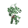
NMR representative
Selection criteria: medoid
NMR ensemble
Conformer selection criteria: structures with the least restraint violations Conformers calculated total number: 50 / Conformers submitted total number: 9
+
About Yorodumi
-
News
-
Feb 9, 2022. New format data for meta-information of EMDB entries
New format data for meta-information of EMDB entries
Version 3 of the EMDB header file is now the official format.
The previous official version 1.9 will be removed from the archive.
In the structure databanks used in Yorodumi, some data are registered as the other names, "COVID-19 virus" and "2019-nCoV". Here are the details of the virus and the list of structure data.
Jan 31, 2019. EMDB accession codes are about to change! (news from PDBe EMDB page)
EMDB accession codes are about to change! (news from PDBe EMDB page)
The allocation of 4 digits for EMDB accession codes will soon come to an end. Whilst these codes will remain in use, new EMDB accession codes will include an additional digit and will expand incrementally as the available range of codes is exhausted. The current 4-digit format prefixed with “EMD-” (i.e. EMD-XXXX) will advance to a 5-digit format (i.e. EMD-XXXXX), and so on. It is currently estimated that the 4-digit codes will be depleted around Spring 2019, at which point the 5-digit format will come into force.
The EM Navigator/Yorodumi systems omit the EMD- prefix.
Related info.:Q: What is EMD? / ID/Accession-code notation in Yorodumi/EM Navigator
Yorodumi is a browser for structure data from EMDB, PDB, SASBDB, etc.
This page is also the successor to EM Navigator detail page, and also detail information page/front-end page for Omokage search.
The word "yorodu" (or yorozu) is an old Japanese word meaning "ten thousand". "mi" (miru) is to see.
Related info.:EMDB / PDB / SASBDB / Comparison of 3 databanks / Yorodumi Search / Aug 31, 2016. New EM Navigator & Yorodumi / Yorodumi Papers / Jmol/JSmol / Function and homology information / Changes in new EM Navigator and Yorodumi
 Movie
Movie Controller


 Open data
 Basic information
 Components
 Keywords
 Function and homology information
 Authors
 United States, 1items
  Citation
 Structure visualization
 Downloads & links
 Other downloads

 PDBj
PDBj

 Assembly
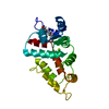
 light scattering

 Mass: 24.305 Da / Num. of mol.: 1 / Source method: obtained synthetically / Formula: Mg
 Sample preparation
 Processing