 Movie
Movie Controller


+ Open data

- Basic information
| Entry | Database: PDB / ID: 7m1n | ||||||
|---|---|---|---|---|---|---|---|
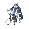
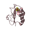
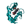
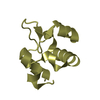
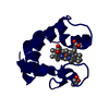
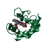
| Title | Wild-type Hydrogenobacter thermophilus ferredoxin 1 | ||||||
 Components Components | Putative ferredoxin | ||||||
 Keywords Keywords | ELECTRON TRANSPORT | ||||||
| Function / homology | : / 4Fe-4S single cluster domain of Ferredoxin I / 4Fe-4S ferredoxin-type iron-sulfur binding domain profile. / 4Fe-4S ferredoxin-type, iron-sulphur binding domain / 4 iron, 4 sulfur cluster binding / metal ion binding / IRON/SULFUR CLUSTER / Putative ferredoxin Function and homology information Function and homology information | ||||||
| Biological species |   Hydrogenobacter thermophilus (bacteria) Hydrogenobacter thermophilus (bacteria) | ||||||
| Method |  X-RAY DIFFRACTION / MOLECULAR REPLACEMENT / Resolution: 2.06 Å X-RAY DIFFRACTION / MOLECULAR REPLACEMENT / Resolution: 2.06 Å | ||||||
 Authors Authors | Steindel, P.S. / Li, B. / Elliott, S.J. | ||||||
| Funding support |  United States, 1items United States, 1items
| ||||||
 Citation Citation | Journal: Acs Catalysis / Year: 2021 Title: Maximizing (Electro)catalytic CO2 Reduction with a Ferredoxin-Based Reduction Potential Gradient Authors: Li, B. / Steindel, P. / Haddad, N. / Elliott, S.J. | ||||||
| History |
|
- Structure visualization
| Structure viewer | Molecule: MolmilJmol/JSmol |
|---|
- Downloads & links
-Download
| PDBx/mmCIF format | 7m1n.cif.gz | 98.9 KB | Display | PDBx/mmCIF format |
|---|---|---|---|---|
| PDB format | pdb7m1n.ent.gz | 73.7 KB | Display | PDB format |
| PDBx/mmJSON format | 7m1n.json.gz | Tree view | PDBx/mmJSON format | |
| Others |  Other downloads Other downloads |
-Validation report
| Arichive directory | https://data.pdbj.org/pub/pdb/validation_reports/m1/7m1nftp://data.pdbj.org/pub/pdb/validation_reports/m1/7m1n | HTTPS FTP |
|---|
-Related structure data
| Related structure data |  7m1oC  1iqzS S: Starting model for refinement C: citing same article ( |
|---|---|
| Similar structure data |


-Links
 PDBj
PDBj


- Assembly
| Deposited unit | 
| |||||||||||||||
|---|---|---|---|---|---|---|---|---|---|---|---|---|---|---|---|---|
| 1 | 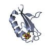
| |||||||||||||||
| 2 | 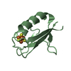
| |||||||||||||||
| Unit cell |
| |||||||||||||||
| Components on special symmetry positions |
|
-Components
| #1: Protein | Mass: 7953.935 Da / Num. of mol.: 2 Source method: isolated from a genetically manipulated source Source: (gene. exp.) Hydrogenobacter thermophilus (bacteria)Gene: fdx1 / Production host: #2: Chemical |   Mass: 351.640 Da / Num. of mol.: 2 / Source method: obtained synthetically / Formula: Fe4S4 / Feature type: SUBJECT OF INVESTIGATION Mass: 351.640 Da / Num. of mol.: 2 / Source method: obtained synthetically / Formula: Fe4S4 / Feature type: SUBJECT OF INVESTIGATION#3: Water | ChemComp-HOH / |  Mass: 18.015 Da / Num. of mol.: 134 / Source method: isolated from a natural source / Formula: H2O Mass: 18.015 Da / Num. of mol.: 134 / Source method: isolated from a natural source / Formula: H2OHas ligand of interest | Y | |
|---|
-Experimental details
-Experiment
| Experiment | Method: X-RAY DIFFRACTION / Number of used crystals: 1 |
|---|
- Sample preparation
| Crystal | Density Matthews: 2.06 Å3/Da / Density % sol: 40.33 % |
|---|---|
| Crystal grow | Temperature: 289 K / Method: vapor diffusion, hanging drop / Details: 79% saturated ammonium sulfate, 0.1 M MES (pH 6.5) |
-Data collection
| Diffraction | Mean temperature: 100 K / Serial crystal experiment: N |
|---|---|
| Diffraction source | Source: ROTATING ANODE / Type: BRUKER AXS MICROSTAR / Wavelength: 1.54 Å |
| Detector | Type: Bruker Platinum 135 / Detector: CCD / Date: Nov 16, 2015 |
| Radiation | Protocol: SINGLE WAVELENGTH / Monochromatic (M) / Laue (L): M / Scattering type: x-ray |
| Radiation wavelength | Wavelength: 1.54 Å / Relative weight: 1 |
| Reflection | Resolution: 2.06→28 Å / Num. obs: 15621 / % possible obs: 98.9 % / Redundancy: 6.6 % / Biso Wilson estimate: 17.0650310623 Å2 / CC1/2: 0.98 / Net I/σ(I): 2.2 |
| Reflection shell | Resolution: 2.06→2.13 Å / Redundancy: 2.1 % / Num. unique obs: 2921 / CC1/2: 0.76 / % possible all: 88.9 |
- Processing
| Software |
| ||||||||||||||||||||||||||||||||||||||||||||||||||||||||||||||||||||||||||||||||||||
|---|---|---|---|---|---|---|---|---|---|---|---|---|---|---|---|---|---|---|---|---|---|---|---|---|---|---|---|---|---|---|---|---|---|---|---|---|---|---|---|---|---|---|---|---|---|---|---|---|---|---|---|---|---|---|---|---|---|---|---|---|---|---|---|---|---|---|---|---|---|---|---|---|---|---|---|---|---|---|---|---|---|---|---|---|---|
| Refinement | Method to determine structure: MOLECULAR REPLACEMENT Starting model: 1IQZ Resolution: 2.06→28 Å / SU ML: 0.192028364882 / Cross valid method: FREE R-VALUE / σ(F): 1.34748017906 / Phase error: 21.5958856637 / Stereochemistry target values: GeoStd + Monomer Library
| ||||||||||||||||||||||||||||||||||||||||||||||||||||||||||||||||||||||||||||||||||||
| Solvent computation | Shrinkage radii: 0.9 Å / VDW probe radii: 1.11 Å / Solvent model: FLAT BULK SOLVENT MODEL | ||||||||||||||||||||||||||||||||||||||||||||||||||||||||||||||||||||||||||||||||||||
| Displacement parameters | Biso mean: 29.1412151389 Å2 | ||||||||||||||||||||||||||||||||||||||||||||||||||||||||||||||||||||||||||||||||||||
| Refinement step | Cycle: LAST / Resolution: 2.06→28 Å
| ||||||||||||||||||||||||||||||||||||||||||||||||||||||||||||||||||||||||||||||||||||
| Refine LS restraints |
| ||||||||||||||||||||||||||||||||||||||||||||||||||||||||||||||||||||||||||||||||||||
| LS refinement shell |
|
