 Movie
Movie Controller

[English] 日本語
 Yorodumi
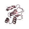
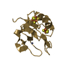
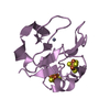
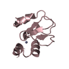
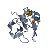
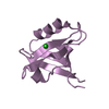
- PDB-1iqz: OXIDIZED [4Fe-4S] FERREDOXIN FROM BACILLUS THERMOPROTEOLYTICUS (F... -
+ Open data

- Basic information
| Entry | Database: PDB / ID: 1iqz | |||||||||
|---|---|---|---|---|---|---|---|---|---|---|
| Title | OXIDIZED [4Fe-4S] FERREDOXIN FROM BACILLUS THERMOPROTEOLYTICUS (FORM I) | |||||||||
 Components Components | Ferredoxin | |||||||||
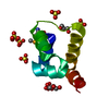
 Keywords Keywords | ELECTRON TRANSPORT / FERREDOXIN / IRON-SULFER PROTEIN / ULTLAHIGH RESOLUTION ANALYSIS / GEOMETRY OF [4FE-4S] CLUSTER | |||||||||
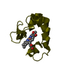
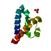
| Function / homology |  Function and homology information Function and homology information4 iron, 4 sulfur cluster binding / electron transfer activity / iron ion binding Similarity search - Function | |||||||||
| Biological species |  | |||||||||
| Method |  X-RAY DIFFRACTION / SYNCHROTRON / FOURIER SYNTHESIS / Resolution: 0.92 Å X-RAY DIFFRACTION / SYNCHROTRON / FOURIER SYNTHESIS / Resolution: 0.92 Å | |||||||||
 Authors Authors | Fukuyama, K. / Okada, T. / Kakuta, Y. / Takahashi, Y. | |||||||||
 Citation Citation | Journal: J.Mol.Biol. / Year: 2002 Title: Atomic resolution structures of oxidized [4Fe-4S] ferredoxin from Bacillus thermoproteolyticus in two crystal forms: systematic distortion of [4Fe-4S] cluster in the protein. Authors: Fukuyama, K. / Okada, T. / Kakuta, Y. / Takahashi, Y. #1: Journal: J.Mol.Biol. / Year: 1989Title: Structure of [4Fe-4S] ferredoxin from Bacillus thermoproteolyticus refined at 2.3 A resolution. Structural comparisons of bacterial ferredoxins. Authors: Fukuyama, K. / Matsubara, H. / Tsukihara, T. / Katsube, Y. #2: Journal: J.Mol.Biol. / Year: 1988Title: Tertiary structure of Bacillus thermoproteolyticus [4Fe-4S] ferredoxin. Evolutionary implications for bacterial ferredoxins. Authors: Fukuyama, K. / Nagahara, Y. / Tsukihara, T. / Katsube, Y. / Hase, T. / Matsubara, H. | |||||||||
| History |
|
- Structure visualization
| Structure viewer | Molecule: MolmilJmol/JSmol |
|---|
- Downloads & links
-Download
| PDBx/mmCIF format | 1iqz.cif.gz | 52.5 KB | Display | PDBx/mmCIF format |
|---|---|---|---|---|
| PDB format | pdb1iqz.ent.gz | 37.2 KB | Display | PDB format |
| PDBx/mmJSON format | 1iqz.json.gz | Tree view | PDBx/mmJSON format | |
| Others |  Other downloads Other downloads |
-Validation report
| Arichive directory | https://data.pdbj.org/pub/pdb/validation_reports/iq/1iqzftp://data.pdbj.org/pub/pdb/validation_reports/iq/1iqz | HTTPS FTP |
|---|
-Related structure data
| Related structure data |  1ir0C  2fxb C: citing same article ( S: Starting model for refinement |
|---|---|
| Similar structure data |

-Links
 PDBj
PDBj


- Assembly
| Deposited unit | 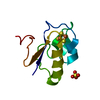
| ||||||||
|---|---|---|---|---|---|---|---|---|---|
| 1 |
| ||||||||
| Unit cell |
| ||||||||
| Components on special symmetry positions |
|
-Components
| #1: Protein | Mass: 8773.595 Da / Num. of mol.: 1 Source method: isolated from a genetically manipulated source Source: (gene. exp.) Plasmid: pET21a(+) / Production host: |
|---|---|
| #2: Chemical | ChemComp-SO4 /   Mass: 96.063 Da / Num. of mol.: 1 / Source method: obtained synthetically / Formula: SO4 Mass: 96.063 Da / Num. of mol.: 1 / Source method: obtained synthetically / Formula: SO4 |
| #3: Chemical | ChemComp-SF4 /   Mass: 351.640 Da / Num. of mol.: 1 / Source method: obtained synthetically / Formula: Fe4S4 Mass: 351.640 Da / Num. of mol.: 1 / Source method: obtained synthetically / Formula: Fe4S4 |
| #4: Water | ChemComp-HOH /  Mass: 18.015 Da / Num. of mol.: 166 / Source method: isolated from a natural source / Formula: H2O Mass: 18.015 Da / Num. of mol.: 166 / Source method: isolated from a natural source / Formula: H2O |
-Experimental details
-Experiment
| Experiment | Method: X-RAY DIFFRACTION / Number of used crystals: 1 |
|---|
- Sample preparation
| Crystal | Density Matthews: 2.24 Å3/Da / Density % sol: 44.7 % | ||||||||||||||||||
|---|---|---|---|---|---|---|---|---|---|---|---|---|---|---|---|---|---|---|---|
| Crystal grow | Temperature: 277 K / Method: microdialysis / pH: 6.5 Details: ammonium sulfate, MES, pH 6.5, MICRODIALYSIS, temperature 277.0K | ||||||||||||||||||
| Crystal grow | *PLUS PH range low: 7.2 / PH range high: 6.5 | ||||||||||||||||||
| Components of the solutions | *PLUS
|
-Data collection
| Diffraction | Mean temperature: 100 K |
|---|---|
| Diffraction source | Source: SYNCHROTRON / Site: SPring-8  / Beamline: BL40B2 / Wavelength: 0.7 / Wavelength: 0.7 Å / Beamline: BL40B2 / Wavelength: 0.7 / Wavelength: 0.7 Å |
| Detector | Type: ADSC QUANTUM 4 / Detector: CCD / Date: Jun 6, 2000 / Details: BENT CYLINDER |
| Radiation | Monochromator: FIXED-EXIT DOUBLE CRYSTAL / Protocol: SINGLE WAVELENGTH / Monochromatic (M) / Laue (L): M / Scattering type: x-ray |
| Radiation wavelength | Wavelength: 0.7 Å / Relative weight: 1 |
| Reflection | Resolution: 0.92→13.5 Å / Num. all: 54415 / Num. obs: 54415 / % possible obs: 99.5 % / Observed criterion σ(F): 0 / Observed criterion σ(I): 0 / Redundancy: 4.9 % / Rmerge(I) obs: 0.074 / Net I/σ(I): 6.5 |
| Reflection shell | Resolution: 0.92→0.96 Å / Redundancy: 3.6 % / Rmerge(I) obs: 0.219 / Mean I/σ(I) obs: 2.7 / % possible all: 99.5 |
| Reflection | *PLUS Num. measured all: 265846 |
| Reflection shell | *PLUS % possible obs: 99.5 % |
- Processing
| Software |
| |||||||||||||||||||||||||||||||||
|---|---|---|---|---|---|---|---|---|---|---|---|---|---|---|---|---|---|---|---|---|---|---|---|---|---|---|---|---|---|---|---|---|---|---|
| Refinement | Method to determine structure: FOURIER SYNTHESIS Starting model: 2FXB 2fxb Resolution: 0.92→7 Å / Num. parameters: 7323 / Num. restraintsaints: 8708 / Cross valid method: FREE R / σ(F): 0 / Stereochemistry target values: ENGH AND HUBER
| |||||||||||||||||||||||||||||||||
| Refine analyze | Num. disordered residues: 6 / Occupancy sum hydrogen: 486 / Occupancy sum non hydrogen: 766 | |||||||||||||||||||||||||||||||||
| Refinement step | Cycle: LAST / Resolution: 0.92→7 Å
| |||||||||||||||||||||||||||||||||
| Refine LS restraints |
| |||||||||||||||||||||||||||||||||
| Software | *PLUS Name: SHELXL-97 / Classification: refinement | |||||||||||||||||||||||||||||||||
| Refinement | *PLUS σ(F): 0 / % reflection Rfree: 9.5 % / Rfactor all: 0.098 / Rfactor Rfree: 0.11 | |||||||||||||||||||||||||||||||||
| Solvent computation | *PLUS | |||||||||||||||||||||||||||||||||
| Displacement parameters | *PLUS | |||||||||||||||||||||||||||||||||
| Refine LS restraints | *PLUS
|
