 Movie
Movie Controller

[English] 日本語
 Yorodumi
- PDB-7lyy: Crystal Structure of Ebola zaire Envelope glycoprotein GP in comp... -
+ Open data

- Basic information
| Entry | Database: PDB / ID: 7lyy | ||||||
|---|---|---|---|---|---|---|---|
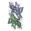
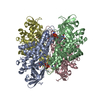
| Title | Crystal Structure of Ebola zaire Envelope glycoprotein GP in complex with compound ARN0075164 | ||||||
 Components Components |
| ||||||
 Keywords Keywords | VIRAL PROTEIN / SSGCID / Envelope glycoprotein / Zaire ebolavirus / Structural Genomics / Seattle Structural Genomics Center for Infectious Disease | ||||||
| Function / homology |  Function and homology information Function and homology informationsymbiont-mediated killing of host cell / host cell endoplasmic reticulum / viral budding from plasma membrane / clathrin-dependent endocytosis of virus by host cell / symbiont-mediated-mediated suppression of host tetherin activity / entry receptor-mediated virion attachment to host cell / host cell cytoplasm / symbiont-mediated suppression of host innate immune response / membrane raft / fusion of virus membrane with host endosome membrane ...symbiont-mediated killing of host cell / host cell endoplasmic reticulum / viral budding from plasma membrane / clathrin-dependent endocytosis of virus by host cell / symbiont-mediated-mediated suppression of host tetherin activity / entry receptor-mediated virion attachment to host cell / host cell cytoplasm / symbiont-mediated suppression of host innate immune response / membrane raft / fusion of virus membrane with host endosome membrane / viral envelope / symbiont entry into host cell / lipid binding / host cell plasma membrane / virion membrane / extracellular region / identical protein binding Similarity search - Function | ||||||
| Biological species |   Zaire ebolavirus Zaire ebolavirus | ||||||
| Method |  X-RAY DIFFRACTION / SYNCHROTRON / MOLECULAR REPLACEMENT / Resolution: 2.75 Å X-RAY DIFFRACTION / SYNCHROTRON / MOLECULAR REPLACEMENT / Resolution: 2.75 Å | ||||||
 Authors Authors | Seattle Structural Genomics Center for Infectious Disease (SSGCID) | ||||||
 Citation Citation | Journal: to be published Title: Crystal Structure of Ebola zaire Envelope glycoprotein GP in complex with compound ARN0075164 Authors: Abendroth, J. / Horanyi, P.S. / Lorimer, D.D. / Edwards, T.E. | ||||||
| History |
|
- Structure visualization
| Structure viewer | Molecule: MolmilJmol/JSmol |
|---|
- Downloads & links
-Download
| PDBx/mmCIF format | 7lyy.cif.gz | 166 KB | Display | PDBx/mmCIF format |
|---|---|---|---|---|
| PDB format | pdb7lyy.ent.gz | 128.3 KB | Display | PDB format |
| PDBx/mmJSON format | 7lyy.json.gz | Tree view | PDBx/mmJSON format | |
| Others |  Other downloads Other downloads |
-Validation report
| Arichive directory | https://data.pdbj.org/pub/pdb/validation_reports/ly/7lyyftp://data.pdbj.org/pub/pdb/validation_reports/ly/7lyy | HTTPS FTP |
|---|
-Related structure data
| Related structure data |  6f5uS S: Starting model for refinement |
|---|---|
| Similar structure data |


-Links
 PDBj
PDBj

- Assembly
| Deposited unit | 
| ||||||||||
|---|---|---|---|---|---|---|---|---|---|---|---|
| 1 | 
| ||||||||||
| Unit cell |
|
-Components
-Protein , 2 types, 2 molecules AB
| #1: Protein | Mass: 32224.197 Da / Num. of mol.: 1 / Fragment: proteolyzed EbzaA.19907.a.HE11 N-terminal domain / Mutation: T42A Source method: isolated from a genetically manipulated source Source: (gene. exp.) Zaire ebolavirus / Strain: Mayinga-76 / Gene: GP / Plasmid: EbzaA..19907.a.HE11 / Production host:  |
|---|---|
| #2: Protein | Mass: 18922.320 Da / Num. of mol.: 1 / Fragment: proteolyzed EbzaA.19907.a.HE11 C-terminal domain / Mutation: H613A Source method: isolated from a genetically manipulated source Source: (gene. exp.) Zaire ebolavirus / Strain: Mayinga-76 / Gene: GP / Plasmid: EbzaA.19907.a.HE11 / Production host: |
-Sugars , 2 types, 5 molecules 
| #3: Polysaccharide | alpha-D-mannopyranose-(1-6)-beta-D-mannopyranose-(1-4)-2-acetamido-2-deoxy-beta-D-glucopyranose-(1- ...alpha-D-mannopyranose-(1-6)-beta-D-mannopyranose-(1-4)-2-acetamido-2-deoxy-beta-D-glucopyranose-(1-4)-2-acetamido-2-deoxy-beta-D-glucopyranose Source method: isolated from a genetically manipulated source |
|---|---|
| #4: Sugar | ChemComp-NAG /  Type: D-saccharide, beta linking / Mass: 221.208 Da / Num. of mol.: 4 Type: D-saccharide, beta linking / Mass: 221.208 Da / Num. of mol.: 4Source method: isolated from a genetically manipulated source Formula: C8H15NO6 |
-Non-polymers , 3 types, 82 molecules 


| #5: Chemical |  Mass: 62.068 Da / Num. of mol.: 3 / Source method: obtained synthetically / Formula: C2H6O2 Mass: 62.068 Da / Num. of mol.: 3 / Source method: obtained synthetically / Formula: C2H6O2#6: Chemical | ChemComp-YPM / ( |  Mass: 428.609 Da / Num. of mol.: 1 / Source method: obtained synthetically / Formula: C29H36N2O Mass: 428.609 Da / Num. of mol.: 1 / Source method: obtained synthetically / Formula: C29H36N2O#7: Water | ChemComp-HOH / | Mass: 18.015 Da / Num. of mol.: 78 / Source method: isolated from a natural source / Formula: H2O |
|---|
-Details
| Has ligand of interest | Y |
|---|---|
| Has protein modification | Y |
| Sequence details | Residues in the C-terminal portion of chain A were difficult to identify, and so were modeled as ...Residues in the C-terminal portion of chain A were difficult to identify, and so were modeled as UNK. The actual sequence for chain A is: ETGRSIPLGV |
-Experimental details
-Experiment
| Experiment | Method: X-RAY DIFFRACTION / Number of used crystals: 1 |
|---|
- Sample preparation
| Crystal | Density Matthews: 3.38 Å3/Da / Density % sol: 63.7 % |
|---|---|
| Crystal grow | Temperature: 287 K / Method: vapor diffusion, sitting drop / pH: 5 Details: Molecular Dimensions / Calibre Proplex screen e2: 100mM sodium citrate pH 5.0, 8% (w/V) PEG 8000: EbzaA.19907.a.HE11.PD38351 at 10.61 mg/ml + 1mM compound AN75164/BSI110991: tray 313199e2, ...Details: Molecular Dimensions / Calibre Proplex screen e2: 100mM sodium citrate pH 5.0, 8% (w/V) PEG 8000: EbzaA.19907.a.HE11.PD38351 at 10.61 mg/ml + 1mM compound AN75164/BSI110991: tray 313199e2, cryo: 20% EG + 5mM compound: puck puk2-8 |
-Data collection
| Diffraction | Mean temperature: 100 K / Serial crystal experiment: N |
|---|---|
| Diffraction source | Source: SYNCHROTRON / Site: APS  / Beamline: 21-ID-F / Wavelength: 0.97872 Å / Beamline: 21-ID-F / Wavelength: 0.97872 Å |
| Detector | Type: RAYONIX MX-300 / Detector: CCD / Date: Feb 8, 2021 / Details: Beryllium Lenses |
| Radiation | Monochromator: Diamond [111] / Protocol: SINGLE WAVELENGTH / Monochromatic (M) / Laue (L): M / Scattering type: x-ray |
| Radiation wavelength | Wavelength: 0.97872 Å / Relative weight: 1 |
| Reflection | Resolution: 2.75→50 Å / Num. obs: 20588 / % possible obs: 99.9 % / Redundancy: 5.511 % / Biso Wilson estimate: 66.76 Å2 / Rmerge(I) obs: 0.06 / Net I/σ(I): 18.6 |
| Reflection shell | Resolution: 2.75→2.82 Å / Redundancy: 5.66 % / Rmerge(I) obs: 0.597 / Mean I/σ(I) obs: 2.84 / Num. unique obs: 1504 / % possible all: 100 |
- Processing
| Software |
| |||||||||||||||||||||||||||||||||||||||||||||||||||||||||||||||||||||||||||||||||||||||||||||||||||||||||||||||||||||||||||||||||||||||||||||||||||||||||||||||||||||||||||||||||||||||||||||||||||||||||||||||||||||||||||||||||||||||||||||||||||||||||||||||||||||||||||||||||||||||||||||||||||||||||||||||||||||||||||||||||||||
|---|---|---|---|---|---|---|---|---|---|---|---|---|---|---|---|---|---|---|---|---|---|---|---|---|---|---|---|---|---|---|---|---|---|---|---|---|---|---|---|---|---|---|---|---|---|---|---|---|---|---|---|---|---|---|---|---|---|---|---|---|---|---|---|---|---|---|---|---|---|---|---|---|---|---|---|---|---|---|---|---|---|---|---|---|---|---|---|---|---|---|---|---|---|---|---|---|---|---|---|---|---|---|---|---|---|---|---|---|---|---|---|---|---|---|---|---|---|---|---|---|---|---|---|---|---|---|---|---|---|---|---|---|---|---|---|---|---|---|---|---|---|---|---|---|---|---|---|---|---|---|---|---|---|---|---|---|---|---|---|---|---|---|---|---|---|---|---|---|---|---|---|---|---|---|---|---|---|---|---|---|---|---|---|---|---|---|---|---|---|---|---|---|---|---|---|---|---|---|---|---|---|---|---|---|---|---|---|---|---|---|---|---|---|---|---|---|---|---|---|---|---|---|---|---|---|---|---|---|---|---|---|---|---|---|---|---|---|---|---|---|---|---|---|---|---|---|---|---|---|---|---|---|---|---|---|---|---|---|---|---|---|---|---|---|---|---|---|---|---|---|---|---|---|---|---|---|---|---|---|---|---|---|---|---|---|---|---|---|---|---|---|---|---|---|---|---|---|---|---|---|---|---|---|---|---|---|---|---|---|---|---|---|---|---|---|---|---|---|---|---|---|---|---|---|---|---|
| Refinement | Method to determine structure: MOLECULAR REPLACEMENT Starting model: PDB ENTRY 6F5U Resolution: 2.75→47.16 Å / SU ML: 0.349 / Cross valid method: FREE R-VALUE / σ(F): 1.35 / Phase error: 23.117 Stereochemistry target values: GEOSTD + MONOMER LIBRARY + CDL V1.2
| |||||||||||||||||||||||||||||||||||||||||||||||||||||||||||||||||||||||||||||||||||||||||||||||||||||||||||||||||||||||||||||||||||||||||||||||||||||||||||||||||||||||||||||||||||||||||||||||||||||||||||||||||||||||||||||||||||||||||||||||||||||||||||||||||||||||||||||||||||||||||||||||||||||||||||||||||||||||||||||||||||||
| Solvent computation | Shrinkage radii: 0.9 Å / VDW probe radii: 1.11 Å / Solvent model: FLAT BULK SOLVENT MODEL | |||||||||||||||||||||||||||||||||||||||||||||||||||||||||||||||||||||||||||||||||||||||||||||||||||||||||||||||||||||||||||||||||||||||||||||||||||||||||||||||||||||||||||||||||||||||||||||||||||||||||||||||||||||||||||||||||||||||||||||||||||||||||||||||||||||||||||||||||||||||||||||||||||||||||||||||||||||||||||||||||||||
| Displacement parameters | Biso mean: 79.7 Å2 | |||||||||||||||||||||||||||||||||||||||||||||||||||||||||||||||||||||||||||||||||||||||||||||||||||||||||||||||||||||||||||||||||||||||||||||||||||||||||||||||||||||||||||||||||||||||||||||||||||||||||||||||||||||||||||||||||||||||||||||||||||||||||||||||||||||||||||||||||||||||||||||||||||||||||||||||||||||||||||||||||||||
| Refinement step | Cycle: LAST / Resolution: 2.75→47.16 Å
| |||||||||||||||||||||||||||||||||||||||||||||||||||||||||||||||||||||||||||||||||||||||||||||||||||||||||||||||||||||||||||||||||||||||||||||||||||||||||||||||||||||||||||||||||||||||||||||||||||||||||||||||||||||||||||||||||||||||||||||||||||||||||||||||||||||||||||||||||||||||||||||||||||||||||||||||||||||||||||||||||||||
| Refine LS restraints |
| |||||||||||||||||||||||||||||||||||||||||||||||||||||||||||||||||||||||||||||||||||||||||||||||||||||||||||||||||||||||||||||||||||||||||||||||||||||||||||||||||||||||||||||||||||||||||||||||||||||||||||||||||||||||||||||||||||||||||||||||||||||||||||||||||||||||||||||||||||||||||||||||||||||||||||||||||||||||||||||||||||||
| LS refinement shell |
| |||||||||||||||||||||||||||||||||||||||||||||||||||||||||||||||||||||||||||||||||||||||||||||||||||||||||||||||||||||||||||||||||||||||||||||||||||||||||||||||||||||||||||||||||||||||||||||||||||||||||||||||||||||||||||||||||||||||||||||||||||||||||||||||||||||||||||||||||||||||||||||||||||||||||||||||||||||||||||||||||||||
| Refinement TLS params. | Method: refined / Refine-ID: X-RAY DIFFRACTION
| |||||||||||||||||||||||||||||||||||||||||||||||||||||||||||||||||||||||||||||||||||||||||||||||||||||||||||||||||||||||||||||||||||||||||||||||||||||||||||||||||||||||||||||||||||||||||||||||||||||||||||||||||||||||||||||||||||||||||||||||||||||||||||||||||||||||||||||||||||||||||||||||||||||||||||||||||||||||||||||||||||||
| Refinement TLS group |
|
