 Movie
Movie Controller


+ Open data

- Basic information
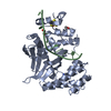
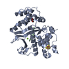
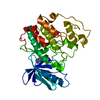
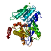
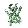
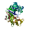
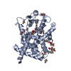
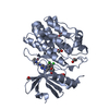
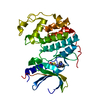
| Entry | Database: PDB / ID: 7lw7 | |||||||||
|---|---|---|---|---|---|---|---|---|---|---|
| Title | Human Exonuclease 5 crystal structure | |||||||||
 Components Components | Exonuclease V | |||||||||
 Keywords Keywords | HYDROLASE | |||||||||
| Function / homology |  Function and homology information Function and homology informationsingle-stranded DNA 5'-3' DNA exonuclease activity / single-stranded DNA 3'-5' DNA exonuclease activity / interstrand cross-link repair / 4 iron, 4 sulfur cluster binding / Hydrolases; Acting on ester bonds / DNA binding / nucleoplasm / metal ion binding / nucleus / cytosol Similarity search - Function | |||||||||
| Biological species |  Homo sapiens (human) Homo sapiens (human) | |||||||||
| Method |  X-RAY DIFFRACTION / SYNCHROTRON / SAD / Resolution: 2.5 Å X-RAY DIFFRACTION / SYNCHROTRON / SAD / Resolution: 2.5 Å | |||||||||
 Authors Authors | Tsai, C.L. / Tainer, J.A. | |||||||||
| Funding support |  United States, 2items United States, 2items
| |||||||||
 Citation Citation | Journal: Mol.Cell / Year: 2021 Title: EXO5-DNA structure and BLM interactions direct DNA resection critical for ATR-dependent replication restart. Authors: Hambarde, S. / Tsai, C.L. / Pandita, R.K. / Bacolla, A. / Maitra, A. / Charaka, V. / Hunt, C.R. / Kumar, R. / Limbo, O. / Le Meur, R. / Chazin, W.J. / Tsutakawa, S.E. / Russell, P. / ...Authors: Hambarde, S. / Tsai, C.L. / Pandita, R.K. / Bacolla, A. / Maitra, A. / Charaka, V. / Hunt, C.R. / Kumar, R. / Limbo, O. / Le Meur, R. / Chazin, W.J. / Tsutakawa, S.E. / Russell, P. / Schlacher, K. / Pandita, T.K. / Tainer, J.A. | |||||||||
| History |
|
- Structure visualization
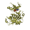
| Structure viewer | Molecule: MolmilJmol/JSmol |
|---|
- Downloads & links
-Download
| PDBx/mmCIF format | 7lw7.cif.gz | 74.2 KB | Display | PDBx/mmCIF format |
|---|---|---|---|---|
| PDB format | pdb7lw7.ent.gz | 52.8 KB | Display | PDB format |
| PDBx/mmJSON format | 7lw7.json.gz | Tree view | PDBx/mmJSON format | |
| Others |  Other downloads Other downloads |
-Validation report
| Arichive directory | https://data.pdbj.org/pub/pdb/validation_reports/lw/7lw7ftp://data.pdbj.org/pub/pdb/validation_reports/lw/7lw7 | HTTPS FTP |
|---|
-Related structure data

-Links
 PDBj
PDBj
- Assembly
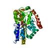
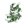
| Deposited unit | 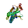
| ||||||||
|---|---|---|---|---|---|---|---|---|---|
| 1 |
| ||||||||
| Unit cell |
|
-Components
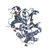
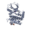
| #1: Protein | Mass: 38856.949 Da / Num. of mol.: 1 / Mutation: G145V Source method: isolated from a genetically manipulated source Source: (gene. exp.) Homo sapiens (human) / Gene: EXO5, C1orf176, DEM1 / Production host:  References: UniProt: Q9H790, Hydrolases; Acting on ester bonds | ||||
|---|---|---|---|---|---|
| #2: Chemical | ChemComp-GOL /   Mass: 92.094 Da / Num. of mol.: 1 / Source method: obtained synthetically / Formula: C3H8O3 Mass: 92.094 Da / Num. of mol.: 1 / Source method: obtained synthetically / Formula: C3H8O3 | ||||
| #3: Chemical | ChemComp-SF4 /   Mass: 351.640 Da / Num. of mol.: 1 / Source method: obtained synthetically / Formula: Fe4S4 / Feature type: SUBJECT OF INVESTIGATION Mass: 351.640 Da / Num. of mol.: 1 / Source method: obtained synthetically / Formula: Fe4S4 / Feature type: SUBJECT OF INVESTIGATION | ||||
| #4: Chemical | ChemComp-EDO /   Mass: 62.068 Da / Num. of mol.: 6 / Source method: obtained synthetically / Formula: C2H6O2 Mass: 62.068 Da / Num. of mol.: 6 / Source method: obtained synthetically / Formula: C2H6O2#5: Water | ChemComp-HOH / |  Mass: 18.015 Da / Num. of mol.: 89 / Source method: isolated from a natural source / Formula: H2O Mass: 18.015 Da / Num. of mol.: 89 / Source method: isolated from a natural source / Formula: H2OHas ligand of interest | Y | |
-Experimental details
-Experiment
| Experiment | Method: X-RAY DIFFRACTION / Number of used crystals: 1 |
|---|
- Sample preparation
| Crystal | Density Matthews: 2.63 Å3/Da / Density % sol: 53.24 % / Description: needle |
|---|---|
| Crystal grow | Temperature: 293 K / Method: vapor diffusion, hanging drop / Details: 20% PEG 3350 and 0.2 M lithium acetate |
-Data collection
| Diffraction | Mean temperature: 100 K / Serial crystal experiment: N | |||||||||||||||||||||||||||
|---|---|---|---|---|---|---|---|---|---|---|---|---|---|---|---|---|---|---|---|---|---|---|---|---|---|---|---|---|
| Diffraction source | Source: SYNCHROTRON / Site: ALS / Beamline: 12.3.1 / Wavelength: 1.1158 Å | |||||||||||||||||||||||||||
| Detector | Type: ADSC QUANTUM 315r / Detector: CCD / Date: Apr 5, 2017 | |||||||||||||||||||||||||||
| Radiation | Protocol: SINGLE WAVELENGTH / Monochromatic (M) / Laue (L): M / Scattering type: x-ray | |||||||||||||||||||||||||||
| Radiation wavelength | Wavelength: 1.1158 Å / Relative weight: 1 | |||||||||||||||||||||||||||
| Reflection | Resolution: 2.5→47.983 Å / Num. obs: 14791 / % possible obs: 100 % / Redundancy: 6.4 % / CC1/2: 0.988 / Rmerge(I) obs: 0.246 / Rpim(I) all: 0.104 / Rrim(I) all: 0.267 / Net I/σ(I): 8.2 | |||||||||||||||||||||||||||
| Reflection shell | Diffraction-ID: 1
|
- Processing
| Software |
| ||||||||||||||||||||||||||||||
|---|---|---|---|---|---|---|---|---|---|---|---|---|---|---|---|---|---|---|---|---|---|---|---|---|---|---|---|---|---|---|---|
| Refinement | Method to determine structure: SAD / Resolution: 2.5→47.983 Å / SU ML: 0.31 / Cross valid method: THROUGHOUT / σ(F): 1.34 / Phase error: 22.51 / Stereochemistry target values: ML
| ||||||||||||||||||||||||||||||
| Solvent computation | Shrinkage radii: 0.9 Å / VDW probe radii: 1.11 Å / Solvent model: FLAT BULK SOLVENT MODEL | ||||||||||||||||||||||||||||||
| Displacement parameters | Biso max: 133.98 Å2 / Biso mean: 41.6189 Å2 / Biso min: 10.75 Å2 | ||||||||||||||||||||||||||||||
| Refinement step | Cycle: final / Resolution: 2.5→47.983 Å
| ||||||||||||||||||||||||||||||
| Refine LS restraints |
| ||||||||||||||||||||||||||||||
| LS refinement shell | Refine-ID: X-RAY DIFFRACTION / Rfactor Rfree error: 0 / % reflection obs: 100 %
|
