 Movie
Movie Controller

[English] 日本語
 Yorodumi
- PDB-7lw0: Structural and Biochemical Insight into Assembly of Molecular Mot... -
+ Open data

- Basic information
| Entry | Database: PDB / ID: 7lw0 | ||||||
|---|---|---|---|---|---|---|---|
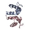
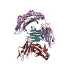
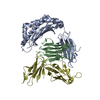
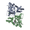
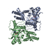
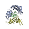
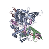
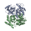
| Title | Structural and Biochemical Insight into Assembly of Molecular Motors Involved in Viral DNA Packaging | ||||||
 Components Components | Terminase small subunit | ||||||
 Keywords Keywords | VIRAL PROTEIN / DNA Packaging / Terminase | ||||||
| Function / homology |  Function and homology information Function and homology informationviral terminase, small subunit / sequence-specific DNA binding, bending / viral DNA genome packaging / Hydrolases; Acting on acid anhydrides; Acting on acid anhydrides to facilitate cellular and subcellular movement / host cell cytoplasm / ATP hydrolysis activity / ATP binding Similarity search - Function | ||||||
| Biological species |  Escherichia phage lambda (virus) Escherichia phage lambda (virus) | ||||||
| Method |  X-RAY DIFFRACTION / SYNCHROTRON / SAD / Resolution: 2.32 Å X-RAY DIFFRACTION / SYNCHROTRON / SAD / Resolution: 2.32 Å | ||||||
 Authors Authors | Ortega, M.E. | ||||||
 Citation Citation | Journal: To Be Published Title: Structural and Biochemical Insight into Assembly of Molecular Motors Involved in Viral DNA Packaging Authors: Ortega, M.E. / Randriamihaja, A. / Rossen, N. / Brannon, J.P. / Marquez, C. / West, R. / Dabbagh, S. / Robles, R. / LeGue, A. | ||||||
| History |
|
- Structure visualization
| Structure viewer | Molecule: MolmilJmol/JSmol |
|---|
- Downloads & links
-Download
| PDBx/mmCIF format | 7lw0.cif.gz | 94.6 KB | Display | PDBx/mmCIF format |
|---|---|---|---|---|
| PDB format | pdb7lw0.ent.gz | 73.8 KB | Display | PDB format |
| PDBx/mmJSON format | 7lw0.json.gz | Tree view | PDBx/mmJSON format | |
| Others |  Other downloads Other downloads |
-Validation report
| Arichive directory | https://data.pdbj.org/pub/pdb/validation_reports/lw/7lw0ftp://data.pdbj.org/pub/pdb/validation_reports/lw/7lw0 | HTTPS FTP |
|---|
-Related structure data

-Links
 PDBj
PDBj

- Assembly
| Deposited unit | 
| ||||||||
|---|---|---|---|---|---|---|---|---|---|
| 1 |
| ||||||||
| Unit cell |
|
-Components
| #1: Protein | Mass: 6264.111 Da / Num. of mol.: 8 Source method: isolated from a genetically manipulated source Source: (gene. exp.) Escherichia phage lambda (virus) / Gene: Nu1, lambdap01 / Production host:  References: UniProt: P03707, Hydrolases; Acting on acid anhydrides; Acting on acid anhydrides to facilitate cellular and subcellular movement Has protein modification | Y | |
|---|
-Experimental details
-Experiment
| Experiment | Method: X-RAY DIFFRACTION / Number of used crystals: 1 |
|---|
- Sample preparation
| Crystal | Density Matthews: 2.04 Å3/Da / Density % sol: 39.68 % |
|---|---|
| Crystal grow | Temperature: 277 K / Method: vapor diffusion, hanging drop Details: isopropanol 5%, 10mM magnesium acetate, tris pH 7.0 |
-Data collection
| Diffraction | Mean temperature: 100 K / Serial crystal experiment: N |
|---|---|
| Diffraction source | Source: SYNCHROTRON / Site: APS  / Beamline: 19-ID / Wavelength: 0.987 Å / Beamline: 19-ID / Wavelength: 0.987 Å |
| Detector | Type: ADSC QUANTUM 315 / Detector: CCD / Date: Feb 10, 2010 |
| Radiation | Protocol: SINGLE WAVELENGTH / Monochromatic (M) / Laue (L): M / Scattering type: x-ray |
| Radiation wavelength | Wavelength: 0.987 Å / Relative weight: 1 |
| Reflection | Resolution: 2.32→34.3 Å / Num. obs: 16731 / % possible obs: 97.5 % / Redundancy: 2.6 % / CC1/2: 0.96 / Net I/σ(I): 11.5 |
| Reflection shell | Resolution: 2.32→2.4 Å / Num. unique obs: 1681 / CC1/2: 0.976 |
- Processing
| Software |
| ||||||||||||||||||||||||||||||||||||||||||||||||||||||||||||
|---|---|---|---|---|---|---|---|---|---|---|---|---|---|---|---|---|---|---|---|---|---|---|---|---|---|---|---|---|---|---|---|---|---|---|---|---|---|---|---|---|---|---|---|---|---|---|---|---|---|---|---|---|---|---|---|---|---|---|---|---|---|
| Refinement | Method to determine structure: SAD / Resolution: 2.32→34.26 Å / Cor.coef. Fo:Fc: 0.961 / Cor.coef. Fo:Fc free: 0.945 / SU B: 2.457 / SU ML: 0.06 / Cross valid method: THROUGHOUT / σ(F): 0 / ESU R: 0.609 / ESU R Free: 0.179 / Stereochemistry target values: MAXIMUM LIKELIHOOD Details: HYDROGENS HAVE BEEN ADDED IN THE RIDING POSITIONS U VALUES : REFINED INDIVIDUALLY
| ||||||||||||||||||||||||||||||||||||||||||||||||||||||||||||
| Solvent computation | Ion probe radii: 0.8 Å / Shrinkage radii: 0.8 Å / VDW probe radii: 1.2 Å / Solvent model: MASK | ||||||||||||||||||||||||||||||||||||||||||||||||||||||||||||
| Displacement parameters | Biso max: 169.45 Å2 / Biso mean: 25.537 Å2 / Biso min: 9.27 Å2
| ||||||||||||||||||||||||||||||||||||||||||||||||||||||||||||
| Refinement step | Cycle: final / Resolution: 2.32→34.26 Å
| ||||||||||||||||||||||||||||||||||||||||||||||||||||||||||||
| Refine LS restraints |
| ||||||||||||||||||||||||||||||||||||||||||||||||||||||||||||
| LS refinement shell | Resolution: 2.32→2.376 Å / Rfactor Rfree error: 0
|
