 Movie
Movie Controller

[English] 日本語
 Yorodumi
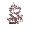
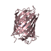
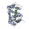
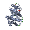
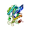
- PDB-7lsm: Crystal structure of E.coli DsbA in complex with bile salt tauroc... -
+ Open data

- Basic information
| Entry | Database: PDB / ID: 7lsm | |||||||||
|---|---|---|---|---|---|---|---|---|---|---|
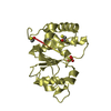
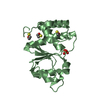
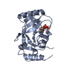
| Title | Crystal structure of E.coli DsbA in complex with bile salt taurocholate | |||||||||
 Components Components | Thiol:disulfide interchange protein DsbA | |||||||||
 Keywords Keywords | OXIDOREDUCTASE / Inhibitor / complex / antibacterial | |||||||||
| Function / homology |  Function and homology information Function and homology informationprotein disulfide isomerase activity / cellular response to antibiotic / Secretion of toxins / protein-disulfide reductase activity / outer membrane-bounded periplasmic space Similarity search - Function | |||||||||
| Biological species |  | |||||||||
| Method |  X-RAY DIFFRACTION / SYNCHROTRON / MOLECULAR REPLACEMENT / Resolution: 1.786 Å X-RAY DIFFRACTION / SYNCHROTRON / MOLECULAR REPLACEMENT / Resolution: 1.786 Å | |||||||||
 Authors Authors | Wang, G. / Heras, B. | |||||||||
| Funding support |  Australia, 2items Australia, 2items
| |||||||||
 Citation Citation | Journal: Chemmedchem / Year: 2022 Title: Selective Binding of Small Molecules to Vibrio cholerae DsbA Offers a Starting Point for the Design of Novel Antibacterials. Authors: Wang, G. / Mohanty, B. / Williams, M.L. / Doak, B.C. / Dhouib, R. / Totsika, M. / McMahon, R.M. / Sharma, G. / Zheng, D. / Bentley, M.R. / Ka-Yan Chin, Y. / Horne, J. / Chalmers, D.K. / Heras, B. / Scanlon, M.J. | |||||||||
| History |
|
- Structure visualization
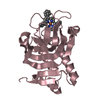
| Structure viewer | Molecule: MolmilJmol/JSmol |
|---|
- Downloads & links
-Download
| PDBx/mmCIF format | 7lsm.cif.gz | 56.8 KB | Display | PDBx/mmCIF format |
|---|---|---|---|---|
| PDB format | pdb7lsm.ent.gz | 37.9 KB | Display | PDB format |
| PDBx/mmJSON format | 7lsm.json.gz | Tree view | PDBx/mmJSON format | |
| Others |  Other downloads Other downloads |
-Validation report
| Arichive directory | https://data.pdbj.org/pub/pdb/validation_reports/ls/7lsmftp://data.pdbj.org/pub/pdb/validation_reports/ls/7lsm | HTTPS FTP |
|---|
-Related structure data
| Related structure data | 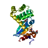 7luiC  1fvkS S: Starting model for refinement C: citing same article ( |
|---|---|
| Similar structure data |

-Links
 PDBj
PDBj

- Assembly
| Deposited unit | 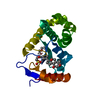
| ||||||||
|---|---|---|---|---|---|---|---|---|---|
| 1 |
| ||||||||
| Unit cell |
|
-Components
| #1: Protein | Mass: 21155.025 Da / Num. of mol.: 1 Source method: isolated from a genetically manipulated source Source: (gene. exp.) Strain: K12 / Gene: dsbA, dsf, ppfA, b3860, JW3832 / Production host: |
|---|---|
| #2: Chemical | ChemComp-PEG /   Mass: 106.120 Da / Num. of mol.: 1 / Source method: obtained synthetically / Formula: C4H10O3 Mass: 106.120 Da / Num. of mol.: 1 / Source method: obtained synthetically / Formula: C4H10O3 |
| #3: Chemical | ChemComp-TCH /   Mass: 515.703 Da / Num. of mol.: 1 / Source method: obtained synthetically / Formula: C26H45NO7S / Feature type: SUBJECT OF INVESTIGATION Mass: 515.703 Da / Num. of mol.: 1 / Source method: obtained synthetically / Formula: C26H45NO7S / Feature type: SUBJECT OF INVESTIGATION |
| #4: Water | ChemComp-HOH /  Mass: 18.015 Da / Num. of mol.: 176 / Source method: isolated from a natural source / Formula: H2O Mass: 18.015 Da / Num. of mol.: 176 / Source method: isolated from a natural source / Formula: H2O |
| Has ligand of interest | Y |
| Has protein modification | Y |
-Experimental details
-Experiment
| Experiment | Method: X-RAY DIFFRACTION / Number of used crystals: 1 |
|---|
- Sample preparation
| Crystal | Density Matthews: 1.85 Å3/Da / Density % sol: 33.4 % |
|---|---|
| Crystal grow | Temperature: 293 K / Method: vapor diffusion, hanging drop Details: 11-13% PEG8000, 5-7.5% glycerol, 1 mM copper(II) chloride, 100 mM sodium cacodylate |
-Data collection
| Diffraction | Mean temperature: 100 K / Serial crystal experiment: N | ||||||||||||||||||||||||||||||
|---|---|---|---|---|---|---|---|---|---|---|---|---|---|---|---|---|---|---|---|---|---|---|---|---|---|---|---|---|---|---|---|
| Diffraction source | Source: SYNCHROTRON / Site: Australian Synchrotron / Beamline: MX1 / Wavelength: 0.95366 Å | ||||||||||||||||||||||||||||||
| Detector | Type: ADSC QUANTUM 210r / Detector: CCD / Date: Sep 22, 2017 | ||||||||||||||||||||||||||||||
| Radiation | Protocol: SINGLE WAVELENGTH / Monochromatic (M) / Laue (L): M / Scattering type: x-ray | ||||||||||||||||||||||||||||||
| Radiation wavelength | Wavelength: 0.95366 Å / Relative weight: 1 | ||||||||||||||||||||||||||||||
| Reflection | Resolution: 1.786→38.3 Å / Num. obs: 15471 / % possible obs: 100 % / Redundancy: 13.9 % / Biso Wilson estimate: 10.18 Å2 / CC1/2: 0.996 / Rmerge(I) obs: 0.126 / Rpim(I) all: 0.035 / Rrim(I) all: 0.13 / Net I/σ(I): 16.2 | ||||||||||||||||||||||||||||||
| Reflection shell | Diffraction-ID: 1
|
- Processing
| Software |
| ||||||||||||||||||||||||||||||
|---|---|---|---|---|---|---|---|---|---|---|---|---|---|---|---|---|---|---|---|---|---|---|---|---|---|---|---|---|---|---|---|
| Refinement | Method to determine structure: MOLECULAR REPLACEMENT Starting model: 1FVK Resolution: 1.786→34.203 Å / SU ML: 0.18 / Cross valid method: THROUGHOUT / σ(F): 1.35 / Phase error: 19.45 / Stereochemistry target values: ML
| ||||||||||||||||||||||||||||||
| Solvent computation | Shrinkage radii: 0.9 Å / VDW probe radii: 1.11 Å / Solvent model: FLAT BULK SOLVENT MODEL | ||||||||||||||||||||||||||||||
| Displacement parameters | Biso max: 53.68 Å2 / Biso mean: 11.2206 Å2 / Biso min: 1.51 Å2 | ||||||||||||||||||||||||||||||
| Refinement step | Cycle: final / Resolution: 1.786→34.203 Å
| ||||||||||||||||||||||||||||||
| Refine LS restraints |
| ||||||||||||||||||||||||||||||
| LS refinement shell | Refine-ID: X-RAY DIFFRACTION / Rfactor Rfree error: 0 / % reflection obs: 100 %
|
