 Movie
Movie Controller

[English] 日本語
 Yorodumi
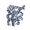
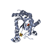
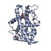
- PDB-7lhu: Crystal structure of adenosine-5'-phosphosulfate reductase from M... -
+ Open data

- Basic information
| Entry | Database: PDB / ID: 7lhu | ||||||
|---|---|---|---|---|---|---|---|
| Title | Crystal structure of adenosine-5'-phosphosulfate reductase from Mycobacterium tuberculosis in a complex with product AMP | ||||||
 Components Components | Phosphoadenosine phosphosulfate reductase | ||||||
 Keywords Keywords | OXIDOREDUCTASE / AMP / adenosine-5'-phosphosulfate reductase / tuberculosis | ||||||
| Function / homology |  Function and homology information Function and homology informationadenylyl-sulfate reductase (thioredoxin) / adenylyl-sulfate reductase (thioredoxin) activity / phosphoadenylyl-sulfate reductase (thioredoxin) activity / : / hydrogen sulfide biosynthetic process / L-cysteine biosynthetic process / 4 iron, 4 sulfur cluster binding / metal ion binding / cytoplasm Similarity search - Function | ||||||
| Biological species |   Mycobacterium tuberculosis (bacteria) Mycobacterium tuberculosis (bacteria) | ||||||
| Method |  X-RAY DIFFRACTION / SYNCHROTRON / MOLECULAR REPLACEMENT / Resolution: 3.09 Å X-RAY DIFFRACTION / SYNCHROTRON / MOLECULAR REPLACEMENT / Resolution: 3.09 Å | ||||||
 Authors Authors | Feliciano, P.R. / Drennan, C.L. | ||||||
| Funding support |  United States, 1items United States, 1items
| ||||||
 Citation Citation | Journal: Acs Omega / Year: 2021 Title: Crystal Structure of the [4Fe-4S] Cluster-Containing Adenosine-5'-phosphosulfate Reductase from Mycobacterium tuberculosis . Authors: Feliciano, P.R. / Carroll, K.S. / Drennan, C.L. | ||||||
| History |
|
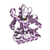
- Structure visualization
| Structure viewer | Molecule: MolmilJmol/JSmol |
|---|
- Downloads & links
-Download
| PDBx/mmCIF format | 7lhu.cif.gz | 180.4 KB | Display | PDBx/mmCIF format |
|---|---|---|---|---|
| PDB format | pdb7lhu.ent.gz | 142 KB | Display | PDB format |
| PDBx/mmJSON format | 7lhu.json.gz | Tree view | PDBx/mmJSON format | |
| Others |  Other downloads Other downloads |
-Validation report
| Arichive directory | https://data.pdbj.org/pub/pdb/validation_reports/lh/7lhuftp://data.pdbj.org/pub/pdb/validation_reports/lh/7lhu | HTTPS FTP |
|---|
-Related structure data
| Related structure data | 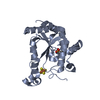 7lhrSC 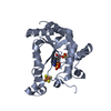 7lhsC S: Starting model for refinement C: citing same article ( |
|---|---|
| Similar structure data |


-Links
 PDBj
PDBj


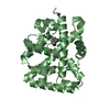
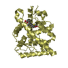
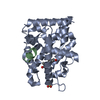
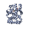
- Assembly
| Deposited unit | 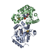
| ||||||||||||||||||||||||||||||||||||||||||||||||||||||||||||||||||||||||||||||||||||||||||
|---|---|---|---|---|---|---|---|---|---|---|---|---|---|---|---|---|---|---|---|---|---|---|---|---|---|---|---|---|---|---|---|---|---|---|---|---|---|---|---|---|---|---|---|---|---|---|---|---|---|---|---|---|---|---|---|---|---|---|---|---|---|---|---|---|---|---|---|---|---|---|---|---|---|---|---|---|---|---|---|---|---|---|---|---|---|---|---|---|---|---|---|
| 1 | 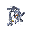
| ||||||||||||||||||||||||||||||||||||||||||||||||||||||||||||||||||||||||||||||||||||||||||
| 2 | 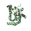
| ||||||||||||||||||||||||||||||||||||||||||||||||||||||||||||||||||||||||||||||||||||||||||
| Unit cell |
| ||||||||||||||||||||||||||||||||||||||||||||||||||||||||||||||||||||||||||||||||||||||||||
| Noncrystallographic symmetry (NCS) | NCS domain:
NCS domain segments: Ens-ID: 1
|