 Movie
Movie Controller


+ Open data

- Basic information
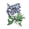
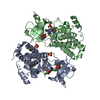
| Entry | Database: PDB / ID: 7lh6 | ||||||
|---|---|---|---|---|---|---|---|
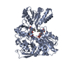
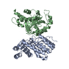
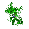
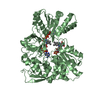
| Title | The structure of Bacteroides plebeius L-galactose dehydrogenase | ||||||
 Components Components | L-galactose dehydrogenase | ||||||
 Keywords Keywords | OXIDOREDUCTASE / dehydrogenase / L-galactose / carbohydrate | ||||||
| Function / homology | L-galactose dehydrogenase activity / L-galactose dehydrogenase-like / Aldo-keto reductase / NADP-dependent oxidoreductase domain / Aldo/keto reductase family / NADP-dependent oxidoreductase domain superfamily / Oxidoreductase, aldo/keto reductase family protein Function and homology information Function and homology information | ||||||
| Biological species |  Bacteroides plebeius (bacteria) Bacteroides plebeius (bacteria) | ||||||
| Method |  X-RAY DIFFRACTION / MOLECULAR REPLACEMENT / Resolution: 2.85 Å X-RAY DIFFRACTION / MOLECULAR REPLACEMENT / Resolution: 2.85 Å | ||||||
 Authors Authors | Robb, C.S. / Pluvinage, B. / Vickers, C. / Boraston, A.B. | ||||||
 Citation Citation | Journal: Nat.Chem.Biol. / Year: 2022 Title: Metabolism of a hybrid algal galactan by members of the human gut microbiome. Authors: Robb, C.S. / Hobbs, J.K. / Pluvinage, B. / Reintjes, G. / Klassen, L. / Monteith, S. / Giljan, G. / Amundsen, C. / Vickers, C. / Hettle, A.G. / Hills, R. / Xing, X. / Montina, T. / Zandberg, ...Authors: Robb, C.S. / Hobbs, J.K. / Pluvinage, B. / Reintjes, G. / Klassen, L. / Monteith, S. / Giljan, G. / Amundsen, C. / Vickers, C. / Hettle, A.G. / Hills, R. / Xing, X. / Montina, T. / Zandberg, W.F. / Abbott, D.W. / Boraston, A.B. | ||||||
| History |
|
- Structure visualization
| Structure viewer | Molecule: MolmilJmol/JSmol |
|---|
- Downloads & links
-Download
| PDBx/mmCIF format | 7lh6.cif.gz | 275.2 KB | Display | PDBx/mmCIF format |
|---|---|---|---|---|
| PDB format | pdb7lh6.ent.gz | 186.4 KB | Display | PDB format |
| PDBx/mmJSON format | 7lh6.json.gz | Tree view | PDBx/mmJSON format | |
| Others |  Other downloads Other downloads |
-Validation report
| Arichive directory | https://data.pdbj.org/pub/pdb/validation_reports/lh/7lh6ftp://data.pdbj.org/pub/pdb/validation_reports/lh/7lh6 | HTTPS FTP |
|---|
-Related structure data
| Related structure data | 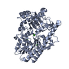 7lhaC 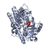 7lj2C  7ljjC  7lk7C  7lnpC 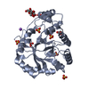 1ynpS S: Starting model for refinement C: citing same article ( |
|---|---|
| Similar structure data |


-Links
 PDBj
PDBj
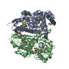
- Assembly
| Deposited unit | 
| ||||||||||||
|---|---|---|---|---|---|---|---|---|---|---|---|---|---|
| 1 |
| ||||||||||||
| Unit cell |
|
-Components
| #1: Protein | Mass: 37096.914 Da / Num. of mol.: 2 Source method: isolated from a genetically manipulated source Source: (gene. exp.) Bacteroides plebeius (bacteria) / Strain: DSM 17135 / JCM 12973 / M2 / Gene: BACPLE_01679 / Production host: #2: Water | ChemComp-HOH / |  Mass: 18.015 Da / Num. of mol.: 17 / Source method: isolated from a natural source / Formula: H2O Mass: 18.015 Da / Num. of mol.: 17 / Source method: isolated from a natural source / Formula: H2O |
|---|
-Experimental details
-Experiment
| Experiment | Method: X-RAY DIFFRACTION / Number of used crystals: 1 |
|---|
- Sample preparation
| Crystal | Density Matthews: 2.04 Å3/Da / Density % sol: 39.61 % |
|---|---|
| Crystal grow | Temperature: 291.15 K / Method: vapor diffusion, hanging drop Details: 0.2 M ammonium citrate dibasic (pH 5.5), 20% (w/v) PEG 3350 |
-Data collection
| Diffraction | Mean temperature: 100 K / Serial crystal experiment: N |
|---|---|
| Diffraction source | Source: ROTATING ANODE / Type: RIGAKU MICROMAX-007 HF / Wavelength: 1.5418 Å |
| Detector | Type: DECTRIS PILATUS 200K / Detector: PIXEL / Date: Jan 21, 2016 |
| Radiation | Protocol: SINGLE WAVELENGTH / Monochromatic (M) / Laue (L): M / Scattering type: x-ray |
| Radiation wavelength | Wavelength: 1.5418 Å / Relative weight: 1 |
| Reflection | Resolution: 2.85→30 Å / Num. obs: 14732 / % possible obs: 98.6 % / Redundancy: 5.8 % / Biso Wilson estimate: 54.55 Å2 / CC1/2: 0.994 / Rmerge(I) obs: 0.106 / Rpim(I) all: 0.046 / Net I/σ(I): 14.7 |
| Reflection shell | Resolution: 2.85→2.9 Å / Redundancy: 3.5 % / Rmerge(I) obs: 0.819 / Mean I/σ(I) obs: 1.7 / Num. unique obs: 704 / CC1/2: 0.82 / Rpim(I) all: 0.478 / % possible all: 95.5 |
- Processing
| Software |
| |||||||||||||||||||||||||||||||||||||||||||||||||||||||||||||||||||||||||||||
|---|---|---|---|---|---|---|---|---|---|---|---|---|---|---|---|---|---|---|---|---|---|---|---|---|---|---|---|---|---|---|---|---|---|---|---|---|---|---|---|---|---|---|---|---|---|---|---|---|---|---|---|---|---|---|---|---|---|---|---|---|---|---|---|---|---|---|---|---|---|---|---|---|---|---|---|---|---|---|
| Refinement | Method to determine structure: MOLECULAR REPLACEMENT Starting model: 1ynp Resolution: 2.85→29.87 Å / SU ML: 0.3823 / Cross valid method: FREE R-VALUE / σ(F): 1.34 / Phase error: 28.6946 / Stereochemistry target values: GeoStd + Monomer Library
| |||||||||||||||||||||||||||||||||||||||||||||||||||||||||||||||||||||||||||||
| Solvent computation | Shrinkage radii: 0.9 Å / VDW probe radii: 1.11 Å / Solvent model: FLAT BULK SOLVENT MODEL | |||||||||||||||||||||||||||||||||||||||||||||||||||||||||||||||||||||||||||||
| Displacement parameters | Biso mean: 51.17 Å2 | |||||||||||||||||||||||||||||||||||||||||||||||||||||||||||||||||||||||||||||
| Refinement step | Cycle: LAST / Resolution: 2.85→29.87 Å
| |||||||||||||||||||||||||||||||||||||||||||||||||||||||||||||||||||||||||||||
| Refine LS restraints |
| |||||||||||||||||||||||||||||||||||||||||||||||||||||||||||||||||||||||||||||
| LS refinement shell |
| |||||||||||||||||||||||||||||||||||||||||||||||||||||||||||||||||||||||||||||
| Refinement TLS params. | Method: refined / Origin x: 46.71393857 Å / Origin y: 40.0006137785 Å / Origin z: 85.9097596471 Å
| |||||||||||||||||||||||||||||||||||||||||||||||||||||||||||||||||||||||||||||
| Refinement TLS group | Selection details: all |
