 Movie
Movie Controller


+ Open data

- Basic information
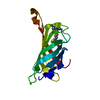
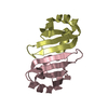
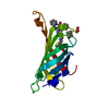
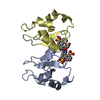
| Entry | Database: PDB / ID: 7lgr | ||||||
|---|---|---|---|---|---|---|---|
| Title | Streptococcus mutans Collagen binding Protein CNM - N2 Domain | ||||||
 Components Components | Collagen-binding adhesin | ||||||
 Keywords Keywords | CELL ADHESION / Streptococcus mutans / CNM / C-terminal N2 Domain / Collagen binding | ||||||
| Function / homology |  Function and homology information Function and homology information | ||||||
| Biological species |  Streptococcus mutans (bacteria) Streptococcus mutans (bacteria) | ||||||
| Method |  X-RAY DIFFRACTION / SYNCHROTRON / MOLECULAR REPLACEMENT / Resolution: 1.6 Å X-RAY DIFFRACTION / SYNCHROTRON / MOLECULAR REPLACEMENT / Resolution: 1.6 Å | ||||||
 Authors Authors | Schormann, N. / Deivanayagam, C. | ||||||
| Funding support |  United States, 1items United States, 1items
| ||||||
 Citation Citation | Journal: To Be Published Title: Streptococcus mutans Collagen binding Protein CNM - Structural and Functional Analysis of the C-terminal N2 Domain Authors: Schormann, N. / Deivanayagam, C. | ||||||
| History |
|
- Structure visualization
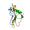
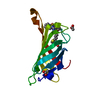
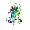
| Structure viewer | Molecule: MolmilJmol/JSmol |
|---|
- Downloads & links
-Download
| PDBx/mmCIF format | 7lgr.cif.gz | 76.4 KB | Display | PDBx/mmCIF format |
|---|---|---|---|---|
| PDB format | pdb7lgr.ent.gz | 54.6 KB | Display | PDB format |
| PDBx/mmJSON format | 7lgr.json.gz | Tree view | PDBx/mmJSON format | |
| Others |  Other downloads Other downloads |
-Validation report
| Arichive directory | https://data.pdbj.org/pub/pdb/validation_reports/lg/7lgrftp://data.pdbj.org/pub/pdb/validation_reports/lg/7lgr | HTTPS FTP |
|---|
-Related structure data
| Related structure data | 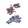 2f6aS S: Starting model for refinement |
|---|---|
| Similar structure data |


-Links
 PDBj
PDBj- Assembly
| Deposited unit | 
| ||||||||
|---|---|---|---|---|---|---|---|---|---|
| 1 |
| ||||||||
| Unit cell |
|
-Components
| #1: Protein | Mass: 19724.160 Da / Num. of mol.: 1 Source method: isolated from a genetically manipulated source Source: (gene. exp.) Streptococcus mutans (bacteria) / Gene: cnm / Plasmid: pET-23d / Production host: |
|---|---|
| #2: Water | ChemComp-HOH /  Mass: 18.015 Da / Num. of mol.: 71 / Source method: isolated from a natural source / Formula: H2O Mass: 18.015 Da / Num. of mol.: 71 / Source method: isolated from a natural source / Formula: H2O |
-Experimental details
-Experiment
| Experiment | Method: X-RAY DIFFRACTION / Number of used crystals: 1 |
|---|
- Sample preparation
| Crystal | Density Matthews: 1.8 Å3/Da / Density % sol: 32 % |
|---|---|
| Crystal grow | Temperature: 293 K / Method: vapor diffusion, hanging drop / pH: 3.5 Details: 1.5M ammonium sulfate, 100mM sodium acetate, pH 4.5 or 100mM citric acid, pH 3.5 |
-Data collection
| Diffraction | Mean temperature: 100 K / Serial crystal experiment: N |
|---|---|
| Diffraction source | Source: SYNCHROTRON / Site: APS / Beamline: 22-ID / Wavelength: 1 Å |
| Detector | Type: MARRESEARCH / Detector: CCD / Date: Dec 14, 2017 |
| Radiation | Protocol: SINGLE WAVELENGTH / Monochromatic (M) / Laue (L): M / Scattering type: x-ray |
| Radiation wavelength | Wavelength: 1 Å / Relative weight: 1 |
| Reflection | Resolution: 1.6→42.05 Å / Num. obs: 19890 / % possible obs: 99.9 % / Redundancy: 10.5 % / Biso Wilson estimate: 22.3 Å2 / CC1/2: 0.999 / Rmerge(I) obs: 0.12 / Rpim(I) all: 0.039 / Rrim(I) all: 0.126 / Net I/σ(I): 13.5 |
| Reflection shell | Resolution: 1.6→1.63 Å / Redundancy: 10.7 % / Rmerge(I) obs: 2.175 / Mean I/σ(I) obs: 1.3 / Num. unique obs: 961 / CC1/2: 0.699 / Rpim(I) all: 0.699 / Rrim(I) all: 2.286 / % possible all: 100 |
- Processing
| Software |
| ||||||||||||||||||||||||||||||||||||||||||||||||||||||||||||||||||||||
|---|---|---|---|---|---|---|---|---|---|---|---|---|---|---|---|---|---|---|---|---|---|---|---|---|---|---|---|---|---|---|---|---|---|---|---|---|---|---|---|---|---|---|---|---|---|---|---|---|---|---|---|---|---|---|---|---|---|---|---|---|---|---|---|---|---|---|---|---|---|---|---|
| Refinement | Method to determine structure: MOLECULAR REPLACEMENT Starting model: 2F6A Resolution: 1.6→42.05 Å / Cor.coef. Fo:Fc: 0.968 / Cor.coef. Fo:Fc free: 0.954 / SU B: 5.59 / SU ML: 0.086 / Cross valid method: THROUGHOUT / σ(F): 0 / ESU R: 0.12 / ESU R Free: 0.098 / Stereochemistry target values: MAXIMUM LIKELIHOOD Details: HYDROGENS HAVE BEEN ADDED IN THE RIDING POSITIONS U VALUES : REFINED INDIVIDUALLY
| ||||||||||||||||||||||||||||||||||||||||||||||||||||||||||||||||||||||
| Solvent computation | Ion probe radii: 0.8 Å / Shrinkage radii: 0.8 Å / VDW probe radii: 1.2 Å / Solvent model: MASK | ||||||||||||||||||||||||||||||||||||||||||||||||||||||||||||||||||||||
| Displacement parameters | Biso max: 83.25 Å2 / Biso mean: 32.8 Å2 / Biso min: 18.72 Å2
| ||||||||||||||||||||||||||||||||||||||||||||||||||||||||||||||||||||||
| Refinement step | Cycle: final / Resolution: 1.6→42.05 Å
| ||||||||||||||||||||||||||||||||||||||||||||||||||||||||||||||||||||||
| Refine LS restraints |
| ||||||||||||||||||||||||||||||||||||||||||||||||||||||||||||||||||||||
| LS refinement shell | Resolution: 1.6→1.657 Å / Rfactor Rfree error: 0 / Total num. of bins used: 10
|
