 Movie
Movie Controller


+ Open data

- Basic information
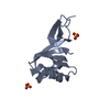
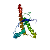
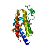
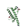
| Entry | Database: PDB / ID: 3eab | ||||||
|---|---|---|---|---|---|---|---|
| Title | Crystal structure of Spastin MIT in complex with ESCRT III | ||||||
 Components Components |
| ||||||
 Keywords Keywords | CELL CYCLE / Spastin / CHMP / MIT / ESCRT / Alternative splicing / ATP-binding / Cytoplasm / Disease mutation / Hereditary spastic paraplegia / Nucleotide-binding / Nucleus / Polymorphism | ||||||
| Function / homology |  Function and homology information Function and homology informationcytokinetic process / microtubule-severing ATPase / microtubule severing ATPase activity / central nervous system neuron axonogenesis / microtubule severing / positive regulation of microtubule depolymerization / MIT domain binding / endoplasmic reticulum tubular network / multivesicular body-lysosome fusion / amphisome membrane ...cytokinetic process / microtubule-severing ATPase / microtubule severing ATPase activity / central nervous system neuron axonogenesis / microtubule severing / positive regulation of microtubule depolymerization / MIT domain binding / endoplasmic reticulum tubular network / multivesicular body-lysosome fusion / amphisome membrane / vesicle fusion with vacuole / ESCRT III complex disassembly / late endosome to lysosome transport / ESCRT III complex / kinetochore microtubule / endosome transport via multivesicular body sorting pathway / cytoskeleton-dependent cytokinesis / nuclear membrane reassembly / membrane coat / mitotic nuclear membrane reassembly / multivesicular body sorting pathway / midbody abscission / Sealing of the nuclear envelope (NE) by ESCRT-III / regulation of centrosome duplication / membrane fission / multivesicular body assembly / late endosome to vacuole transport / multivesicular body membrane / ubiquitin-dependent protein catabolic process via the multivesicular body sorting pathway / anterograde axonal transport / microtubule bundle formation / plasma membrane repair / regulation of mitotic spindle assembly / exit from mitosis / protein hexamerization / mitotic spindle disassembly / mitotic metaphase chromosome alignment / nucleus organization / axonal transport of mitochondrion / positive regulation of cytokinesis / viral budding via host ESCRT complex / mitotic cytokinesis / autophagosome membrane / autophagosome maturation / endoplasmic reticulum to Golgi vesicle-mediated transport / nuclear pore / alpha-tubulin binding / beta-tubulin binding / multivesicular body / axon cytoplasm / lipid droplet / viral budding from plasma membrane / establishment of protein localization / protein homooligomerization / kinetochore / autophagy / spindle / spindle pole / protein transport / cytoplasmic vesicle / midbody / nuclear membrane / microtubule binding / microtubule / endosome / endosome membrane / protein domain specific binding / axon / cell division / lysosomal membrane / centrosome / endoplasmic reticulum membrane / protein-containing complex binding / perinuclear region of cytoplasm / ATP hydrolysis activity / extracellular exosome / nucleoplasm / ATP binding / identical protein binding / nucleus / plasma membrane / cytoplasm / cytosol Similarity search - Function | ||||||
| Biological species |  Homo sapiens (human) Homo sapiens (human) | ||||||
| Method |  X-RAY DIFFRACTION / SYNCHROTRON / MAD / Resolution: 2.5 Å X-RAY DIFFRACTION / SYNCHROTRON / MAD / Resolution: 2.5 Å | ||||||
 Authors Authors | Yang, D. / Rimanchi, N. / Renvoise, B. / Lippincott-Schwartz, J. / Blackstone, C. / Hurley, J.H. | ||||||
 Citation Citation | Journal: Nat.Struct.Mol.Biol. / Year: 2008 Title: Structural basis for midbody targeting of spastin by the ESCRT-III protein CHMP1B. Authors: Yang, D. / Rismanchi, N. / Renvoise, B. / Lippincott-Schwartz, J. / Blackstone, C. / Hurley, J.H. | ||||||
| History |
|
- Structure visualization
| Structure viewer | Molecule: MolmilJmol/JSmol |
|---|
- Downloads & links
-Download
| PDBx/mmCIF format | 3eab.cif.gz | 154 KB | Display | PDBx/mmCIF format |
|---|---|---|---|---|
| PDB format | pdb3eab.ent.gz | 126.8 KB | Display | PDB format |
| PDBx/mmJSON format | 3eab.json.gz | Tree view | PDBx/mmJSON format | |
| Others |  Other downloads Other downloads |
-Validation report
| Arichive directory | https://data.pdbj.org/pub/pdb/validation_reports/ea/3eabftp://data.pdbj.org/pub/pdb/validation_reports/ea/3eab | HTTPS FTP |
|---|
-Related structure data
| Similar structure data |
|---|


-Links
 PDBj
PDBj


- Assembly
| Deposited unit | 
| ||||||||
|---|---|---|---|---|---|---|---|---|---|
| 1 | 
| ||||||||
| 2 | 
| ||||||||
| 3 | 
| ||||||||
| 4 | 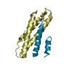
| ||||||||
| 5 | 
| ||||||||
| 6 | 
| ||||||||
| Unit cell |
|
-Components
| #1: Protein | Mass: 10314.937 Da / Num. of mol.: 6 / Fragment: UNP residues 112 to 196 Source method: isolated from a genetically manipulated source Source: (gene. exp.) Homo sapiens (human) / Gene: SPAST, KIAA1083, SPG4 / Production host:  #2: Protein/peptide | Mass: 5420.950 Da / Num. of mol.: 6 / Fragment: UNP residues 145 to 194 Source method: isolated from a genetically manipulated source Source: (gene. exp.) Homo sapiens (human) / Production host: |
|---|
-Experimental details
-Experiment
| Experiment | Method: X-RAY DIFFRACTION / Number of used crystals: 1 |
|---|
- Sample preparation
| Crystal | Density Matthews: 3.76 Å3/Da / Density % sol: 67.28 % |
|---|---|
| Crystal grow | Temperature: 288 K / Method: vapor diffusion, hanging drop / pH: 4.6 Details: 10% PEG 2000 MME, 0.2 M Ammonia Sulfate, 0.1 M Sodium Acetate, pH 4.6, VAPOR DIFFUSION, HANGING DROP, temperature 288K |
-Data collection
| Diffraction | Mean temperature: 100 K | ||||||||||||
|---|---|---|---|---|---|---|---|---|---|---|---|---|---|
| Diffraction source | Source: SYNCHROTRON / Site: APS  / Beamline: 22-ID / Wavelength: 0.97920, 0.97934, 0.97166 / Beamline: 22-ID / Wavelength: 0.97920, 0.97934, 0.97166 | ||||||||||||
| Detector | Type: MAR scanner 300 mm plate / Detector: IMAGE PLATE / Date: Feb 4, 2008 | ||||||||||||
| Radiation | Monochromator: Si 220 / Protocol: MAD / Monochromatic (M) / Laue (L): M / Scattering type: x-ray | ||||||||||||
| Radiation wavelength |
| ||||||||||||
| Reflection | Resolution: 2.3→50 Å / Num. obs: 59615 / % possible obs: 90.9 % / Biso Wilson estimate: 24.5 Å2 | ||||||||||||
| Reflection shell | Resolution: 2.3→2.38 Å / % possible all: 53.3 |
- Processing
| Software |
| |||||||||||||||||||||||||
|---|---|---|---|---|---|---|---|---|---|---|---|---|---|---|---|---|---|---|---|---|---|---|---|---|---|---|
| Refinement | Method to determine structure: MAD / Resolution: 2.5→47.65 Å / Rfactor Rfree error: 0.005 / Data cutoff high absF: 58202.66 / Data cutoff low absF: 0 / Isotropic thermal model: RESTRAINED / Cross valid method: THROUGHOUT / σ(F): 0 / Stereochemistry target values: Engh & Huber
| |||||||||||||||||||||||||
| Solvent computation | Solvent model: FLAT MODEL / Bsol: 43.8832 Å2 / ksol: 0.345367 e/Å3 | |||||||||||||||||||||||||
| Displacement parameters | Biso mean: 53.7 Å2
| |||||||||||||||||||||||||
| Refine analyze |
| |||||||||||||||||||||||||
| Refinement step | Cycle: LAST / Resolution: 2.5→47.65 Å
| |||||||||||||||||||||||||
| Refine LS restraints |
| |||||||||||||||||||||||||
| LS refinement shell | Resolution: 2.5→2.66 Å / Rfactor Rfree error: 0.02 / Total num. of bins used: 6
| |||||||||||||||||||||||||
| Xplor file |
|
