 Movie
Movie Controller


+ Open data

- Basic information
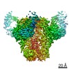
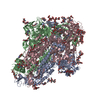
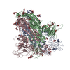
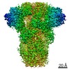
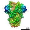
| Entry | Database: PDB / ID: 7lg6 | |||||||||
|---|---|---|---|---|---|---|---|---|---|---|
| Title | BG505 SOSIP.v5.2 in complex with VRC40.01 and RM19R Fabs | |||||||||
 Components Components |
| |||||||||
 Keywords Keywords | VIRAL PROTEIN/IMMUNE SYSTEM / antibody / HIV / SOSIP / Env / VIRAL PROTEIN / VIRAL PROTEIN-IMMUNE SYSTEM complex | |||||||||
| Function / homology |  Function and homology information Function and homology informationsymbiont-mediated perturbation of host defense response / positive regulation of plasma membrane raft polarization / positive regulation of receptor clustering / host cell endosome membrane / clathrin-dependent endocytosis of virus by host cell / viral protein processing / fusion of virus membrane with host plasma membrane / fusion of virus membrane with host endosome membrane / viral envelope / virion attachment to host cell ...symbiont-mediated perturbation of host defense response / positive regulation of plasma membrane raft polarization / positive regulation of receptor clustering / host cell endosome membrane / clathrin-dependent endocytosis of virus by host cell / viral protein processing / fusion of virus membrane with host plasma membrane / fusion of virus membrane with host endosome membrane / viral envelope / virion attachment to host cell / host cell plasma membrane / virion membrane / structural molecule activity / membrane / identical protein binding Similarity search - Function | |||||||||
| Biological species |   Human immunodeficiency virus 1 Human immunodeficiency virus 1 Homo sapiens (human) Homo sapiens (human) | |||||||||
| Method | ELECTRON MICROSCOPY / single particle reconstruction / cryo EM / Resolution: 3.28 Å | |||||||||
 Authors Authors | Cottrell, C.A. / Torres, J.L. / Wu, N.R. / Ward, A.B. | |||||||||
| Funding support |  United States, 2items United States, 2items
| |||||||||
 Citation Citation | Journal: Cell Rep / Year: 2021 Title: Structural basis of glycan276-dependent recognition by HIV-1 broadly neutralizing antibodies. Authors: Christopher A Cottrell / Kartik Manne / Rui Kong / Shuishu Wang / Tongqing Zhou / Gwo-Yu Chuang / Robert J Edwards / Rory Henderson / Katarzyna Janowska / Megan Kopp / Bob C Lin / Mark K ...Authors: Christopher A Cottrell / Kartik Manne / Rui Kong / Shuishu Wang / Tongqing Zhou / Gwo-Yu Chuang / Robert J Edwards / Rory Henderson / Katarzyna Janowska / Megan Kopp / Bob C Lin / Mark K Louder / Adam S Olia / Reda Rawi / Chen-Hsiang Shen / Justin D Taft / Jonathan L Torres / Nelson R Wu / Baoshan Zhang / Nicole A Doria-Rose / Myron S Cohen / Barton F Haynes / Lawrence Shapiro / Andrew B Ward / Priyamvada Acharya / John R Mascola / Peter D Kwong / Abstract: Recognition of N-linked glycan at residue N276 (glycan276) at the periphery of the CD4-binding site (CD4bs) on the HIV-envelope trimer is a formidable challenge for many CD4bs-directed antibodies. To ...Recognition of N-linked glycan at residue N276 (glycan276) at the periphery of the CD4-binding site (CD4bs) on the HIV-envelope trimer is a formidable challenge for many CD4bs-directed antibodies. To understand how this glycan can be recognized, here we isolate two lineages of glycan276-dependent CD4bs antibodies. Antibody CH540-VRC40.01 (named for donor-lineage.clone) neutralizes 81% of a panel of 208 diverse strains, while antibody CH314-VRC33.01 neutralizes 45%. Cryo-electron microscopy (cryo-EM) structures of these two antibodies and 179NC75, a previously identified glycan276-dependent CD4bs antibody, in complex with HIV-envelope trimer reveal substantially different modes of glycan276 recognition. Despite these differences, binding of glycan276-dependent antibodies maintains a glycan276 conformation similar to that observed in the absence of glycan276-binding antibodies. By contrast, glycan276-independent CD4bs antibodies, such as VRC01, displace glycan276 upon binding. These results provide a foundation for understanding antibody recognition of glycan276 and suggest its presence may be crucial for priming immunogens seeking to initiate broad CD4bs recognition. | |||||||||
| History |
|
- Structure visualization
| Movie |
Movie viewer |
|---|---|
| Structure viewer | Molecule: MolmilJmol/JSmol |
- Downloads & links
-Download
| PDBx/mmCIF format | 7lg6.cif.gz | 685.7 KB | Display | PDBx/mmCIF format |
|---|---|---|---|---|
| PDB format | pdb7lg6.ent.gz | 552.9 KB | Display | PDB format |
| PDBx/mmJSON format | 7lg6.json.gz | Tree view | PDBx/mmJSON format | |
| Others |  Other downloads Other downloads |
-Validation report
| Arichive directory | https://data.pdbj.org/pub/pdb/validation_reports/lg/7lg6ftp://data.pdbj.org/pub/pdb/validation_reports/lg/7lg6 | HTTPS FTP |
|---|
-Related structure data
| Related structure data |  23312MC  7ll1C  7ll2C  7llkC M: map data used to model this data C: citing same article ( |
|---|---|
| Similar structure data |


-Links
 PDBj
PDBj


- Assembly
| Deposited unit | 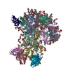
|
|---|---|
| 1 |
|
-Components
-Envelope glycoprotein ... , 2 types, 6 molecules AEFBGI
| #1: Protein | Mass: 53268.371 Da / Num. of mol.: 3 / Mutation: E64K, A73C, A316W, T332N, A501C Source method: isolated from a genetically manipulated source Source: (gene. exp.) Human immunodeficiency virus 1 / Gene: env / Cell line (production host): HEK293F / Production host: Homo sapiens (human) / References: UniProt: Q2N0S6#4: Protein | Mass: 17178.549 Da / Num. of mol.: 3 / Mutation: I559P, A561C, T605C Source method: isolated from a genetically manipulated source Source: (gene. exp.) Human immunodeficiency virus 1 / Gene: env / Cell line (production host): HEK293F / Production host: Homo sapiens (human) / References: UniProt: Q2N0S6 |
|---|
-Antibody , 4 types, 12 molecules HOPLQRDMNCJK
| #2: Antibody | Mass: 24800.186 Da / Num. of mol.: 3 Source method: isolated from a genetically manipulated source Source: (gene. exp.) Homo sapiens (human) / Cell line (production host): HEK293F / Production host: Homo sapiens (human)#3: Antibody | Mass: 23616.271 Da / Num. of mol.: 3 Source method: isolated from a genetically manipulated source Source: (gene. exp.) Homo sapiens (human) / Cell line (production host): HEK293F / Production host: Homo sapiens (human)#5: Antibody | Mass: 23568.156 Da / Num. of mol.: 3 Source method: isolated from a genetically manipulated source Source: (gene. exp.) Homo sapiens (human)#6: Antibody | Mass: 23979.713 Da / Num. of mol.: 3 Source method: isolated from a genetically manipulated source Source: (gene. exp.) Homo sapiens (human) |
|---|
-Sugars , 6 types, 60 molecules 
| #7: Polysaccharide | 2-acetamido-2-deoxy-beta-D-glucopyranose-(1-4)-2-acetamido-2-deoxy-beta-D-glucopyranose Source method: isolated from a genetically manipulated source #8: Polysaccharide | Source method: isolated from a genetically manipulated source #9: Polysaccharide | Source method: isolated from a genetically manipulated source #10: Polysaccharide | Source method: isolated from a genetically manipulated source #11: Polysaccharide | Source method: isolated from a genetically manipulated source #12: Sugar | ChemComp-NAG /  Type: D-saccharide, beta linking / Mass: 221.208 Da / Num. of mol.: 18 Type: D-saccharide, beta linking / Mass: 221.208 Da / Num. of mol.: 18Source method: isolated from a genetically manipulated source Formula: C8H15NO6 |
|---|
-Details
| Has ligand of interest | N |
|---|---|
| Has protein modification | Y |
-Experimental details
-Experiment
| Experiment | Method: ELECTRON MICROSCOPY |
|---|---|
| EM experiment | Aggregation state: PARTICLE / 3D reconstruction method: single particle reconstruction |
- Sample preparation
| Component |
| ||||||||||||||||||||||||||||||
|---|---|---|---|---|---|---|---|---|---|---|---|---|---|---|---|---|---|---|---|---|---|---|---|---|---|---|---|---|---|---|---|
| Molecular weight | Experimental value: NO | ||||||||||||||||||||||||||||||
| Source (natural) |
| ||||||||||||||||||||||||||||||
| Source (recombinant) |
| ||||||||||||||||||||||||||||||
| Buffer solution | pH: 7.4 | ||||||||||||||||||||||||||||||
| Specimen | Embedding applied: NO / Shadowing applied: NO / Staining applied: NO / Vitrification applied: YES | ||||||||||||||||||||||||||||||
| Specimen support | Grid material: COPPER / Grid type: C-flat-1.2/1.3 | ||||||||||||||||||||||||||||||
| Vitrification | Cryogen name: ETHANE |
- Electron microscopy imaging
| Experimental equipment |  Model: Titan Krios / Image courtesy: FEI Company |
|---|---|
| Microscopy | Model: FEI TITAN KRIOS |
| Electron gun | Electron source:  FIELD EMISSION GUN / Accelerating voltage: 300 kV / Illumination mode: FLOOD BEAM FIELD EMISSION GUN / Accelerating voltage: 300 kV / Illumination mode: FLOOD BEAM |
| Electron lens | Mode: BRIGHT FIELD |
| Image recording | Average exposure time: 12.25 sec. / Electron dose: 51.5 e/Å2 / Detector mode: COUNTING / Film or detector model: GATAN K2 SUMMIT (4k x 4k) / Num. of grids imaged: 1 / Num. of real images: 842 |
| Image scans | Movie frames/image: 49 |
- Processing
| EM software |
| ||||||||||||||||||
|---|---|---|---|---|---|---|---|---|---|---|---|---|---|---|---|---|---|---|---|
| CTF correction | Type: PHASE FLIPPING AND AMPLITUDE CORRECTION | ||||||||||||||||||
| Symmetry | Point symmetry: C3 (3 fold cyclic) | ||||||||||||||||||
| 3D reconstruction | Resolution: 3.28 Å / Resolution method: FSC 0.143 CUT-OFF / Num. of particles: 32186 / Symmetry type: POINT | ||||||||||||||||||
| Atomic model building | Protocol: AB INITIO MODEL / Space: REAL / Target criteria: High EMRinger, low Molprobity |
