 Movie
Movie Controller


+ Open data

- Basic information
| Entry | Database: PDB / ID: 7ld5 | ||||||
|---|---|---|---|---|---|---|---|
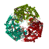
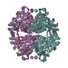
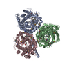
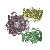
| Title | polynucleotide phosphorylase | ||||||
 Components Components |
| ||||||
 Keywords Keywords | TRANSFERASE/RNA / phosphorylase polymerase RNA decay / RNA BINDING PROTEIN / TRANSFERASE-RNA complex | ||||||
| Function / homology |  Function and homology information Function and homology informationpolyribonucleotide nucleotidyltransferase / polyribonucleotide nucleotidyltransferase activity / mRNA catabolic process / RNA processing / 3'-5'-RNA exonuclease activity / magnesium ion binding / RNA binding / cytosol Similarity search - Function | ||||||
| Biological species |  Mycolicibacterium smegmatis (bacteria) Mycolicibacterium smegmatis (bacteria)synthetic construct (others) | ||||||
| Method | ELECTRON MICROSCOPY / single particle reconstruction / cryo EM / Resolution: 3.07 Å | ||||||
 Authors Authors | Goldgur, Y. / Shuman, S. / De La Cruz, M.J. / Ghosh, S. / Unciuleac, M.-C. | ||||||
| Funding support |  United States, 1items United States, 1items
| ||||||
 Citation Citation | Journal: RNA / Year: 2021 Title: Structure and mechanism of polynucleotide phosphorylase. Authors: Mihaela-Carmen Unciuleac / Shreya Ghosh / M Jason de la Cruz / Yehuda Goldgur / Stewart Shuman / Abstract: Polynucleotide phosphorylase (PNPase) catalyzes stepwise phosphorolysis of the 3'-terminal phosphodiesters of RNA chains to yield nucleoside diphosphate products. In the reverse reaction, PNPase acts ...Polynucleotide phosphorylase (PNPase) catalyzes stepwise phosphorolysis of the 3'-terminal phosphodiesters of RNA chains to yield nucleoside diphosphate products. In the reverse reaction, PNPase acts as a polymerase, using NDPs as substrates to add NMPs to the 3'-OH terminus of RNA chains while expelling inorganic phosphate. The apparent essentiality of PNPase for growth of militates for mycobacterial PNPase as a potential drug target. A cryo-EM structure of PNPase (MsmPNPase) reveals a characteristic ring-shaped homotrimer in which each protomer consists of two RNase PH-like domains and an intervening α-helical module on the inferior surface of the ring. The carboxy-terminal KH and S1 domains, which impart RNA specificity to MsmPNPase, are on the opposite face of the core ring and are conformationally mobile. Single particle reconstructions of MsmPNPase in the act of poly(A) synthesis highlight a 3'-terminal (rA) oligonucleotide and two magnesium ions in the active site and an adenine nucleobase in the central tunnel. We identify amino acids that engage the 3' segment of the RNA chain (Phe68, Arg105, Arg112, Arg430, Arg431) and the two metal ions (Asp526, Asp532, Gln546, Asp548), and we infer those that bind inorganic phosphate (Thr470, Ser471, His435, Lys534). Alanine mutagenesis pinpointed RNA and phosphate contacts as essential (Arg105, Arg431, Lys534, Thr470 + Ser471), important (Arg112, Arg430), or unimportant (Phe68) for PNPase activity. Severe phosphorolysis and polymerase defects accompanying alanine mutations of the enzymic metal ligands suggest a two-metal mechanism of catalysis by MsmPNPase. | ||||||
| History |
|
- Structure visualization
| Movie |
Movie viewer |
|---|---|
| Structure viewer | Molecule: MolmilJmol/JSmol |
- Downloads & links
-Download
| PDBx/mmCIF format | 7ld5.cif.gz | 307.2 KB | Display | PDBx/mmCIF format |
|---|---|---|---|---|
| PDB format | pdb7ld5.ent.gz | 245.7 KB | Display | PDB format |
| PDBx/mmJSON format | 7ld5.json.gz | Tree view | PDBx/mmJSON format | |
| Others |  Other downloads Other downloads |
-Validation report
| Arichive directory | https://data.pdbj.org/pub/pdb/validation_reports/ld/7ld5ftp://data.pdbj.org/pub/pdb/validation_reports/ld/7ld5 | HTTPS FTP |
|---|
-Related structure data
| Related structure data |  23282MC M: map data used to model this data C: citing same article ( |
|---|---|
| Similar structure data |


-Links
 PDBj
PDBj


- Assembly
| Deposited unit | 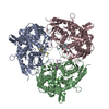
|
|---|---|
| 1 |
|
-Components
| #1: Protein | Mass: 83647.703 Da / Num. of mol.: 3 Source method: isolated from a genetically manipulated source Source: (gene. exp.) Mycolicibacterium smegmatis (bacteria) / Gene: pnp, gpsI, MSMEG_2656, MSMEI_2593 / Production host: References: UniProt: A0QVQ5, polyribonucleotide nucleotidyltransferase #2: RNA chain | Mass: 2917.895 Da / Num. of mol.: 3 / Source method: obtained synthetically / Source: (synth.) synthetic construct (others) #3: Chemical | ChemComp-MG /   Mass: 24.305 Da / Num. of mol.: 6 / Source method: obtained synthetically / Formula: Mg Mass: 24.305 Da / Num. of mol.: 6 / Source method: obtained synthetically / Formula: MgHas ligand of interest | N | Sequence details | Authors state that the exact length of RNA fragment is unknown. | |
|---|
-Experimental details
-Experiment
| Experiment | Method: ELECTRON MICROSCOPY |
|---|---|
| EM experiment | Aggregation state: PARTICLE / 3D reconstruction method: single particle reconstruction |
- Sample preparation
| Component | Name: Polynucleotide phosphorylase complex in poly(A) synthesis Type: COMPLEX / Entity ID: #1-#2 / Source: MULTIPLE SOURCES | ||||||||||||
|---|---|---|---|---|---|---|---|---|---|---|---|---|---|
| Source (natural) |
| ||||||||||||
| Source (recombinant) | Organism: | ||||||||||||
| Buffer solution | pH: 8 | ||||||||||||
| Specimen | Embedding applied: NO / Shadowing applied: NO / Staining applied: NO / Vitrification applied: YES | ||||||||||||
| Vitrification | Cryogen name: ETHANE |
- Electron microscopy imaging
| Experimental equipment |  Model: Titan Krios / Image courtesy: FEI Company |
|---|---|
| Microscopy | Model: FEI TITAN KRIOS |
| Electron gun | Electron source:  FIELD EMISSION GUN / Accelerating voltage: 300 kV / Illumination mode: FLOOD BEAM FIELD EMISSION GUN / Accelerating voltage: 300 kV / Illumination mode: FLOOD BEAM |
| Electron lens | Mode: BRIGHT FIELD |
| Image recording | Electron dose: 53 e/Å2 / Film or detector model: GATAN K3 (6k x 4k) |
- Processing
| Software | Name: PHENIX / Version: 1.17.1_3660: / Classification: refinement | ||||||||||||||||||||||||
|---|---|---|---|---|---|---|---|---|---|---|---|---|---|---|---|---|---|---|---|---|---|---|---|---|---|
| EM software |
| ||||||||||||||||||||||||
| CTF correction | Type: PHASE FLIPPING AND AMPLITUDE CORRECTION | ||||||||||||||||||||||||
| 3D reconstruction | Resolution: 3.07 Å / Resolution method: FSC 0.143 CUT-OFF / Num. of particles: 890249 / Symmetry type: POINT | ||||||||||||||||||||||||
| Atomic model building | Space: REAL | ||||||||||||||||||||||||
| Refine LS restraints |
|
