- PDB-2wit: CRYSTAL STRUCTURE OF THE SODIUM-COUPLED GLYCINE BETAINE SYMPORTER... -
+
Open data
ID or keywords:
Loading...
-
Basic information
Entry
Database: PDB / ID: 2wit
Title
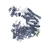
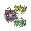
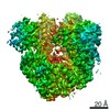
CRYSTAL STRUCTURE OF THE SODIUM-COUPLED GLYCINE BETAINE SYMPORTER BETP FROM CORYNEBACTERIUM GLUTAMICUM WITH BOUND SUBSTRATE
Components
GLYCINE BETAINE TRANSPORTER BETP
Keywords
MEMBRANE PROTEIN / CHEMOSENSOR AND OSMOSENSOR / TRIMER / MEMBRANE / TRANSPORT / CELL MEMBRANE / SECONDARY TRANSPORTER / SODIUM COUPLED TRANSPORT / TRANSMEMBRANE / BETAINE TRANSPORT / HYPEROSMOTIC STRESS
Function / homology
Function and homology information
symporter activity / metal ion binding / identical protein binding / plasma membrane Similarity search - Function
Single alpha-helices involved in coiled-coils or other helix-helix interfaces - #430 / BCCT transporter, conserved site / BCCT family of transporters signature. / BCCT transporter family / BCCT, betaine/carnitine/choline family transporter / Single alpha-helices involved in coiled-coils or other helix-helix interfaces / Up-down Bundle / Mainly Alpha Similarity search - Domain/homology
Type: RAYONIX / Detector: CCD / Date: Feb 12, 2007
Radiation
Monochromator: AL3 / Protocol: SINGLE WAVELENGTH / Monochromatic (M) / Laue (L): M / Scattering type: x-ray
Radiation wavelength
Wavelength: 0.97944 Å / Relative weight: 1
Reflection
Resolution: 3.35→39.47 Å / Num. obs: 37151 / % possible obs: 90.7 % / Observed criterion σ(I): 1 / Redundancy: 18.9 % / Biso Wilson estimate: 27.351 Å2 / Rmerge(I) obs: 0.1 / Net I/σ(I): 23.2
Reflection shell
Resolution: 3.35→3.55 Å / Redundancy: 1.7 % / Rmerge(I) obs: 0.65 / Mean I/σ(I) obs: 1 / % possible all: 44.1
-
Processing
Software
Name
Version
Classification
XDS
datareduction
XSCALE
datascaling
SHELXD
phasing
SHARP
phasing
BUSTER-TNT
2.7.0
refinement
Refinement
Method to determine structure: SAD Starting model: NONE Resolution: 3.35→39.47 Å / Cross valid method: THROUGHOUT / σ(F): 0 Details: HYDROGENS HAVE BEEN ADDED IN THE RIDING POSITIONS. NCS IS PRESENT AND WAS USED IN THE REFINEMENT WITH LOCAL STRUCTURE SIMILARITY RESTRAINTS LSSR. SEE PDB FILE REMARK 3 NCS MODEL RESTRAINT ...Details: HYDROGENS HAVE BEEN ADDED IN THE RIDING POSITIONS. NCS IS PRESENT AND WAS USED IN THE REFINEMENT WITH LOCAL STRUCTURE SIMILARITY RESTRAINTS LSSR. SEE PDB FILE REMARK 3 NCS MODEL RESTRAINT LSSR REMARK 3 TARGET RESTRAINT LSSR SMART, O. S., M. BRANDL, C. FLENSBURG, P. KELLER, W. PACIOREK, C. VONRHEIN, AND G. BRICOGNE. 2008. REFINEMENT WITH LOCAL STRUCTURE SIMILARITY RESTRAINTS LSSR ENABLES EXPLOITATION OF INFORMATION FROM RELATED STRUCTURES AND FACILITATES USE OF NCS. PRESENTED AT THE ANNUAL MEETING OF THE AMERICAN CRYSTALLOGRAPHIC ASSOCIATION. ABSTRACT TP139, KNOXVILLE TN, U.S.A.
Rfactor
Num. reflection
% reflection
Selection details
Rfree
0.2649
1886
5.08 %
RANDOM
Rwork
0.2568
-
-
-
obs
0.2572
37151
90.44 %
-
Displacement parameters
Biso mean: 64.34 Å2
Baniso -1
Baniso -2
Baniso -3
1-
-11.29080734 Å2
0 Å2
0 Å2
2-
-
13.79907882 Å2
0 Å2
3-
-
-
-2.50827148 Å2
Refinement step
Cycle: LAST / Resolution: 3.35→39.47 Å
Protein
Nucleic acid
Ligand
Solvent
Total
Num. atoms
11713
0
24
0
11737
Refine LS restraints
Refine-ID
Type
Number
Restraint function
Weight
X-RAY DIFFRACTION
t_bond_d
12029
0.002
2
X-RAY DIFFRACTION
t_angle_deg
16348
0.342
2
X-RAY DIFFRACTION
t_dihedral_angle_d
1910
9.567
0
X-RAY DIFFRACTION
t_incorr_chiral_ct
X-RAY DIFFRACTION
t_pseud_angle
X-RAY DIFFRACTION
t_trig_c_planes
179
0.001
2
X-RAY DIFFRACTION
t_gen_planes
1802
0.006
5
X-RAY DIFFRACTION
t_it
12008
0.526
50
X-RAY DIFFRACTION
t_nbd
43
0.063
5
X-RAY DIFFRACTION
t_omega_torsion
X-RAY DIFFRACTION
t_other_torsion
X-RAY DIFFRACTION
t_improper_torsion
X-RAY DIFFRACTION
t_chiral_improper_torsion
X-RAY DIFFRACTION
t_sum_occupancies
X-RAY DIFFRACTION
t_utility_distance
X-RAY DIFFRACTION
t_utility_angle
X-RAY DIFFRACTION
t_utility_torsion
X-RAY DIFFRACTION
t_ideal_dist_contact
LS refinement shell
Resolution: 3.35→3.55 Å / Total num. of bins used: 9
Rfactor
Num. reflection
% reflection
Rfree
0.2395
140
4.72 %
Rwork
0.2413
2827
-
all
0.2412
2967
-
obs
-
-
90.44 %
+
About Yorodumi
-
News
-
Feb 9, 2022. New format data for meta-information of EMDB entries
New format data for meta-information of EMDB entries
Version 3 of the EMDB header file is now the official format.
The previous official version 1.9 will be removed from the archive.
In the structure databanks used in Yorodumi, some data are registered as the other names, "COVID-19 virus" and "2019-nCoV". Here are the details of the virus and the list of structure data.
Jan 31, 2019. EMDB accession codes are about to change! (news from PDBe EMDB page)
EMDB accession codes are about to change! (news from PDBe EMDB page)
The allocation of 4 digits for EMDB accession codes will soon come to an end. Whilst these codes will remain in use, new EMDB accession codes will include an additional digit and will expand incrementally as the available range of codes is exhausted. The current 4-digit format prefixed with “EMD-” (i.e. EMD-XXXX) will advance to a 5-digit format (i.e. EMD-XXXXX), and so on. It is currently estimated that the 4-digit codes will be depleted around Spring 2019, at which point the 5-digit format will come into force.
The EM Navigator/Yorodumi systems omit the EMD- prefix.
Related info.:Q: What is EMD? / ID/Accession-code notation in Yorodumi/EM Navigator
Yorodumi is a browser for structure data from EMDB, PDB, SASBDB, etc.
This page is also the successor to EM Navigator detail page, and also detail information page/front-end page for Omokage search.
The word "yorodu" (or yorozu) is an old Japanese word meaning "ten thousand". "mi" (miru) is to see.
Related info.:EMDB / PDB / SASBDB / Comparison of 3 databanks / Yorodumi Search / Aug 31, 2016. New EM Navigator & Yorodumi / Yorodumi Papers / Jmol/JSmol / Function and homology information / Changes in new EM Navigator and Yorodumi
 Movie
Movie Controller

 Yorodumi
 Open data
 Basic information
 Components
 Keywords
 Function and homology information
 CORYNEBACTERIUM GLUTAMICUM (bacteria)
 X-RAY DIFFRACTION /
  Authors
 Citation
 Structure visualization
 Downloads & links
 Other downloads

 PDBj
PDBj Assembly
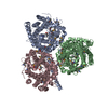

 Mass: 118.154 Da / Num. of mol.: 3 / Source method: obtained synthetically / Formula: C5H12NO2
 Sample preparation
 / Beamline: X10SA / Wavelength: 0.97944
  Processing