 Movie
Movie Controller

[English] 日本語
 Yorodumi
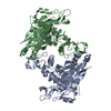
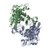
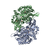
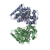
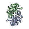
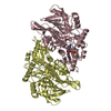
- PDB-7l4i: Crystal structure of a substrate-trapping variant of PPM1H phosphatase -
+ Open data

- Basic information
| Entry | Database: PDB / ID: 7l4i | ||||||
|---|---|---|---|---|---|---|---|
| Title | Crystal structure of a substrate-trapping variant of PPM1H phosphatase | ||||||
 Components Components | Protein phosphatase 1H | ||||||
 Keywords Keywords | HYDROLASE / Rab GTPase / LRRK2 kinase / PP2C/PPM family / membrane trafficking | ||||||
| Function / homology |  Function and homology information Function and homology information[pyruvate dehydrogenase (acetyl-transferring)]-phosphatase activity / protein-serine/threonine phosphatase / phosphoprotein phosphatase activity / glutamatergic synapse / signal transduction / mitochondrion / nucleoplasm / identical protein binding / nucleus / cytoplasm Similarity search - Function | ||||||
| Biological species |  Homo sapiens (human) Homo sapiens (human) | ||||||
| Method |  X-RAY DIFFRACTION / SYNCHROTRON / MOLECULAR REPLACEMENT / Resolution: 2.58 Å X-RAY DIFFRACTION / SYNCHROTRON / MOLECULAR REPLACEMENT / Resolution: 2.58 Å | ||||||
 Authors Authors | Khan, A.R. / Waschbusch, D. | ||||||
 Citation Citation | Journal: Embo Rep. / Year: 2021 Title: Structural basis for the specificity of PPM1H phosphatase for Rab GTPases. Authors: Waschbusch, D. / Berndsen, K. / Lis, P. / Knebel, A. / Lam, Y.P. / Alessi, D.R. / Khan, A.R. | ||||||
| History |
|
- Structure visualization
| Structure viewer | Molecule: MolmilJmol/JSmol |
|---|
- Downloads & links
-Download
| PDBx/mmCIF format | 7l4i.cif.gz | 328.1 KB | Display | PDBx/mmCIF format |
|---|---|---|---|---|
| PDB format | pdb7l4i.ent.gz | 265.5 KB | Display | PDB format |
| PDBx/mmJSON format | 7l4i.json.gz | Tree view | PDBx/mmJSON format | |
| Others |  Other downloads Other downloads |
-Validation report
| Summary document | 7l4i_validation.pdf.gz | 439.4 KB | Display | wwPDB validaton report |
|---|---|---|---|---|
| Full document | 7l4i_full_validation.pdf.gz | 449.7 KB | Display | |
| Data in XML | 7l4i_validation.xml.gz | 29.6 KB | Display | |
| Data in CIF | 7l4i_validation.cif.gz | 40.3 KB | Display | |
| Arichive directory | https://data.pdbj.org/pub/pdb/validation_reports/l4/7l4iftp://data.pdbj.org/pub/pdb/validation_reports/l4/7l4i | HTTPS FTP |
-Related structure data
| Related structure data |  7kprC  7l4jC  7n0zC  6b67S S: Starting model for refinement C: citing same article ( |
|---|---|
| Similar structure data |


-Links
 PDBj
PDBj- Assembly
| Deposited unit | 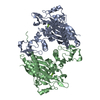
| ||||||||
|---|---|---|---|---|---|---|---|---|---|
| 1 |
| ||||||||
| Unit cell |
|
-Components
| #1: Protein | Mass: 50317.965 Da / Num. of mol.: 2 / Mutation: D288A, C56A Source method: isolated from a genetically manipulated source Source: (gene. exp.) Homo sapiens (human) / Gene: PPM1H, ARHCL1, KIAA1157, URCC2 / Production host:  References: UniProt: Q9ULR3, protein-serine/threonine phosphatase #2: Chemical | ChemComp-MG /   Mass: 24.305 Da / Num. of mol.: 4 / Source method: obtained synthetically / Formula: Mg Mass: 24.305 Da / Num. of mol.: 4 / Source method: obtained synthetically / Formula: Mg#3: Water | ChemComp-HOH / |  Mass: 18.015 Da / Num. of mol.: 91 / Source method: isolated from a natural source / Formula: H2O Mass: 18.015 Da / Num. of mol.: 91 / Source method: isolated from a natural source / Formula: H2OHas ligand of interest | N | |
|---|
-Experimental details
-Experiment
| Experiment | Method: X-RAY DIFFRACTION / Number of used crystals: 1 |
|---|
- Sample preparation
| Crystal | Density Matthews: 2.64 Å3/Da / Density % sol: 53.5 % |
|---|---|
| Crystal grow | Temperature: 298 K / Method: vapor diffusion / Details: 30% PEG 1500 |
-Data collection
| Diffraction | Mean temperature: 100 K / Serial crystal experiment: N |
|---|---|
| Diffraction source | Source: SYNCHROTRON / Site: NSLS-II  / Beamline: 17-ID-2 / Wavelength: 0.979279 Å / Beamline: 17-ID-2 / Wavelength: 0.979279 Å |
| Detector | Type: DECTRIS EIGER X 16M / Detector: PIXEL / Date: Sep 30, 2019 |
| Radiation | Monochromator: 0.979279 / Protocol: SINGLE WAVELENGTH / Monochromatic (M) / Laue (L): M / Scattering type: x-ray |
| Radiation wavelength | Wavelength: 0.979279 Å / Relative weight: 1 |
| Reflection | Resolution: 2.58→28.97 Å / Num. obs: 34086 / % possible obs: 99.5 % / Redundancy: 6.6 % / CC1/2: 0.998 / Rmerge(I) obs: 0.093 / Rpim(I) all: 0.039 / Rrim(I) all: 0.101 / Net I/σ(I): 13.6 / Num. measured all: 224906 |
| Reflection shell | Resolution: 2.58→2.7 Å / Redundancy: 6.5 % / Rmerge(I) obs: 0.651 / Num. unique obs: 3998 / CC1/2: 0.879 / Rpim(I) all: 0.272 / Rrim(I) all: 0.707 / % possible all: 96.9 |
- Processing
| Software |
| ||||||||||||||||||||||||||||||||||||||||||||||||||||||||||||||||||||||||
|---|---|---|---|---|---|---|---|---|---|---|---|---|---|---|---|---|---|---|---|---|---|---|---|---|---|---|---|---|---|---|---|---|---|---|---|---|---|---|---|---|---|---|---|---|---|---|---|---|---|---|---|---|---|---|---|---|---|---|---|---|---|---|---|---|---|---|---|---|---|---|---|---|---|
| Refinement | Method to determine structure: MOLECULAR REPLACEMENT Starting model: 6b67 Resolution: 2.58→28.97 Å / SU ML: 0.29 / Cross valid method: THROUGHOUT / σ(F): 1.35 / Phase error: 26.08 / Stereochemistry target values: ML
| ||||||||||||||||||||||||||||||||||||||||||||||||||||||||||||||||||||||||
| Solvent computation | Shrinkage radii: 0.9 Å / VDW probe radii: 1.11 Å / Solvent model: FLAT BULK SOLVENT MODEL | ||||||||||||||||||||||||||||||||||||||||||||||||||||||||||||||||||||||||
| Displacement parameters | Biso max: 132.78 Å2 / Biso mean: 57.6578 Å2 / Biso min: 27.45 Å2 | ||||||||||||||||||||||||||||||||||||||||||||||||||||||||||||||||||||||||
| Refinement step | Cycle: final / Resolution: 2.58→28.97 Å
| ||||||||||||||||||||||||||||||||||||||||||||||||||||||||||||||||||||||||
| LS refinement shell | Refine-ID: X-RAY DIFFRACTION / Rfactor Rfree error: 0
| ||||||||||||||||||||||||||||||||||||||||||||||||||||||||||||||||||||||||
| Refinement TLS params. | Method: refined / Origin x: 16.9335 Å / Origin y: 60.984 Å / Origin z: 95.9169 Å
| ||||||||||||||||||||||||||||||||||||||||||||||||||||||||||||||||||||||||
| Refinement TLS group |
|
