 Movie
Movie Controller


+ Open data

- Basic information
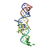
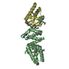
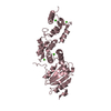
| Entry | Database: PDB / ID: 7l0z | ||||||
|---|---|---|---|---|---|---|---|
| Title | Spinach variant bound to DFHBI-1T | ||||||
 Components Components | RNA (69-MER) | ||||||
 Keywords Keywords | RNA / fluorescent / aptamer | ||||||
| Function / homology | Chem-2ZY / : / PHOSPHATE ION / SPERMINE / RNA / RNA (> 10) Function and homology information Function and homology information | ||||||
| Biological species | synthetic construct (others) | ||||||
| Method |  X-RAY DIFFRACTION / SYNCHROTRON / MOLECULAR REPLACEMENT / Resolution: 2.1 Å X-RAY DIFFRACTION / SYNCHROTRON / MOLECULAR REPLACEMENT / Resolution: 2.1 Å | ||||||
 Authors Authors | Truong, L. / Trachman, R.J. / Ferre-D'Amare, A.R. | ||||||
 Citation Citation | Journal: Rna / Year: 2021 Title: Fluorogenic aptamers resolve the flexibility of RNA junctions using orientation-dependent FRET. Authors: Jeng, S.C.Y. / Trachman III, R.J. / Weissenboeck, F. / Truong, L. / Link, K.A. / Jepsen, M.D.E. / Knutson, J.R. / Andersen, E.S. / Ferre-D'Amare, A.R. / Unrau, P.J. | ||||||
| History |
|
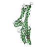
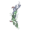
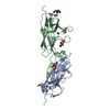
- Structure visualization
| Structure viewer | Molecule: MolmilJmol/JSmol |
|---|
- Downloads & links
-Download
| PDBx/mmCIF format | 7l0z.cif.gz | 112.6 KB | Display | PDBx/mmCIF format |
|---|---|---|---|---|
| PDB format | pdb7l0z.ent.gz | 72 KB | Display | PDB format |
| PDBx/mmJSON format | 7l0z.json.gz | Tree view | PDBx/mmJSON format | |
| Others |  Other downloads Other downloads |
-Validation report
| Arichive directory | https://data.pdbj.org/pub/pdb/validation_reports/l0/7l0zftp://data.pdbj.org/pub/pdb/validation_reports/l0/7l0z | HTTPS FTP |
|---|
-Related structure data
| Related structure data |  6up0C  5ob3S S: Starting model for refinement C: citing same article ( |
|---|---|
| Similar structure data |


-Links
 PDBj
PDBj


- Assembly
| Deposited unit | 
| ||||||||||||
|---|---|---|---|---|---|---|---|---|---|---|---|---|---|
| 1 |
| ||||||||||||
| Unit cell |
| ||||||||||||
| Components on special symmetry positions |
|
-Components
-RNA chain , 1 types, 1 molecules G
| #1: RNA chain | Mass: 22373.242 Da / Num. of mol.: 1 / Source method: obtained synthetically / Source: (synth.) synthetic construct (others) |
|---|
-Non-polymers , 7 types, 65 molecules 


| #2: Chemical | ChemComp-2ZY / ( Mass: 320.215 Da / Num. of mol.: 1 / Source method: obtained synthetically / Formula: C13H9F5N2O2 Mass: 320.215 Da / Num. of mol.: 1 / Source method: obtained synthetically / Formula: C13H9F5N2O2 | ||||||||||
|---|---|---|---|---|---|---|---|---|---|---|---|
| #3: Chemical |  Mass: 202.340 Da / Num. of mol.: 3 / Source method: obtained synthetically / Formula: C10H26N4 Mass: 202.340 Da / Num. of mol.: 3 / Source method: obtained synthetically / Formula: C10H26N4#4: Chemical | ChemComp-MG / |  Mass: 24.305 Da / Num. of mol.: 1 / Source method: obtained synthetically / Formula: Mg Mass: 24.305 Da / Num. of mol.: 1 / Source method: obtained synthetically / Formula: Mg#5: Chemical |  Mass: 94.971 Da / Num. of mol.: 2 / Source method: obtained synthetically / Formula: PO4 Mass: 94.971 Da / Num. of mol.: 2 / Source method: obtained synthetically / Formula: PO4#6: Chemical |  Mass: 22.990 Da / Num. of mol.: 2 / Source method: obtained synthetically / Formula: Na Mass: 22.990 Da / Num. of mol.: 2 / Source method: obtained synthetically / Formula: Na#7: Chemical |  Mass: 39.098 Da / Num. of mol.: 2 / Source method: obtained synthetically / Formula: K Mass: 39.098 Da / Num. of mol.: 2 / Source method: obtained synthetically / Formula: K#8: Water | ChemComp-HOH / | Mass: 18.015 Da / Num. of mol.: 54 / Source method: isolated from a natural source / Formula: H2O |
-Details
| Has ligand of interest | N |
|---|
-Experimental details
-Experiment
| Experiment | Method: X-RAY DIFFRACTION / Number of used crystals: 1 |
|---|
- Sample preparation
| Crystal | Density Matthews: 2.51 Å3/Da / Density % sol: 50.94 % |
|---|---|
| Crystal grow | Temperature: 294 K / Method: vapor diffusion, sitting drop / pH: 7.5 / Details: 40 mM Tris-HCl pH 7.5, 100 mM KCl, 1 mM MgCl2 |
-Data collection
| Diffraction | Mean temperature: 100 K / Serial crystal experiment: N |
|---|---|
| Diffraction source | Source: SYNCHROTRON / Site: APS  / Beamline: 24-ID-C / Wavelength: 1.105 Å / Beamline: 24-ID-C / Wavelength: 1.105 Å |
| Detector | Type: DECTRIS PILATUS3 S 6M / Detector: PIXEL / Date: Nov 17, 2018 |
| Radiation | Protocol: SINGLE WAVELENGTH / Monochromatic (M) / Laue (L): M / Scattering type: x-ray |
| Radiation wavelength | Wavelength: 1.105 Å / Relative weight: 1 |
| Reflection | Resolution: 2.01→75.26 Å / Num. obs: 14648 / % possible obs: 98.7 % / Redundancy: 5.3 % / Biso Wilson estimate: 46.31 Å2 / CC1/2: 0.999 / Net I/σ(I): 13.5 |
| Reflection shell | Resolution: 2.01→2.07 Å / Num. unique obs: 1167 / CC1/2: 0.627 / % possible all: 92.1 |
- Processing
| Software |
| ||||||||||||||||||||||||||||||||||||||||||||||||||||||||||||||||||||||
|---|---|---|---|---|---|---|---|---|---|---|---|---|---|---|---|---|---|---|---|---|---|---|---|---|---|---|---|---|---|---|---|---|---|---|---|---|---|---|---|---|---|---|---|---|---|---|---|---|---|---|---|---|---|---|---|---|---|---|---|---|---|---|---|---|---|---|---|---|---|---|---|
| Refinement | Method to determine structure: MOLECULAR REPLACEMENT Starting model: 5OB3 Resolution: 2.1→75.26 Å / SU ML: 0.2582 / Cross valid method: FREE R-VALUE / σ(F): 1.36 / Phase error: 25.8783
| ||||||||||||||||||||||||||||||||||||||||||||||||||||||||||||||||||||||
| Solvent computation | Shrinkage radii: 0.9 Å / VDW probe radii: 1.11 Å | ||||||||||||||||||||||||||||||||||||||||||||||||||||||||||||||||||||||
| Displacement parameters | Biso mean: 66.07 Å2 | ||||||||||||||||||||||||||||||||||||||||||||||||||||||||||||||||||||||
| Refinement step | Cycle: LAST / Resolution: 2.1→75.26 Å
| ||||||||||||||||||||||||||||||||||||||||||||||||||||||||||||||||||||||
| Refine LS restraints |
| ||||||||||||||||||||||||||||||||||||||||||||||||||||||||||||||||||||||
| LS refinement shell |
| ||||||||||||||||||||||||||||||||||||||||||||||||||||||||||||||||||||||
| Refinement TLS params. | Method: refined / Origin x: -36.8152928881 Å / Origin y: 12.3705808217 Å / Origin z: -0.479627498632 Å
| ||||||||||||||||||||||||||||||||||||||||||||||||||||||||||||||||||||||
| Refinement TLS group | Selection details: all |
