 Movie
Movie Controller

[English] 日本語
 Yorodumi
- PDB-7knj: C1B domain of Protein kinase C in complex with Phorbol ester and ... -
+ Open data

- Basic information
| Entry | Database: PDB / ID: 7knj | ||||||
|---|---|---|---|---|---|---|---|
| Title | C1B domain of Protein kinase C in complex with Phorbol ester and Phosphatidylcholine | ||||||
 Components Components | Protein kinase C delta type | ||||||
 Keywords Keywords | LIPID BINDING PROTEIN / C1 / lipid-binding / phorbol ester binding / Zn2+ finger | ||||||
| Function / homology |  Function and homology information Function and homology informationprotein kinase C / diacylglycerol-dependent serine/threonine kinase activity / response to glucose / intracellular signal transduction / apoptotic process / perinuclear region of cytoplasm / zinc ion binding / ATP binding / nucleus / plasma membrane Similarity search - Function | ||||||
| Biological species |  | ||||||
| Method |  X-RAY DIFFRACTION / MOLECULAR REPLACEMENT / Resolution: 1.57 Å X-RAY DIFFRACTION / MOLECULAR REPLACEMENT / Resolution: 1.57 Å | ||||||
 Authors Authors | Katti, S.S. / Krieger, I. | ||||||
| Funding support |  United States, 1items United States, 1items
| ||||||
 Citation Citation | Journal: Nat Commun / Year: 2022 Title: Structural anatomy of Protein Kinase C C1 domain interactions with diacylglycerol and other agonists. Authors: Katti, S.S. / Krieger, I.V. / Ann, J. / Lee, J. / Sacchettini, J.C. / Igumenova, T.I. | ||||||
| History |
|
- Structure visualization
| Structure viewer | Molecule: MolmilJmol/JSmol |
|---|
- Downloads & links
-Download
| PDBx/mmCIF format | 7knj.cif.gz | 30.9 KB | Display | PDBx/mmCIF format |
|---|---|---|---|---|
| PDB format | pdb7knj.ent.gz | 17 KB | Display | PDB format |
| PDBx/mmJSON format | 7knj.json.gz | Tree view | PDBx/mmJSON format | |
| Others |  Other downloads Other downloads |
-Validation report
| Arichive directory | https://data.pdbj.org/pub/pdb/validation_reports/kn/7knjftp://data.pdbj.org/pub/pdb/validation_reports/kn/7knj | HTTPS FTP |
|---|
-Related structure data
| Related structure data | 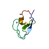 7kndC 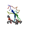 7ko6C  7l92C 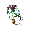 7lcbC 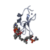 7leoC 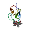 7lf3C 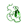 1ptqS S: Starting model for refinement C: citing same article ( |
|---|---|
| Similar structure data |
-Links
 PDBj
PDBj


- Assembly
| Deposited unit | 
| ||||||||
|---|---|---|---|---|---|---|---|---|---|
| 1 |
| ||||||||
| Unit cell |
| ||||||||
| Components on special symmetry positions |
|
-Components
| #1: Protein | Mass: 6097.217 Da / Num. of mol.: 1 / Fragment: C1B domain Source method: isolated from a genetically manipulated source Source: (gene. exp.)  | ||||||||
|---|---|---|---|---|---|---|---|---|---|
| #2: Chemical |   Mass: 482.568 Da / Num. of mol.: 2 / Source method: obtained synthetically / Formula: C22H45NO8P / Feature type: SUBJECT OF INVESTIGATION Mass: 482.568 Da / Num. of mol.: 2 / Source method: obtained synthetically / Formula: C22H45NO8P / Feature type: SUBJECT OF INVESTIGATION#3: Chemical |   Mass: 65.409 Da / Num. of mol.: 2 / Source method: obtained synthetically / Formula: Zn Mass: 65.409 Da / Num. of mol.: 2 / Source method: obtained synthetically / Formula: Zn#4: Chemical | ChemComp-WTS / | 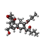  Mass: 504.612 Da / Num. of mol.: 1 / Source method: obtained synthetically / Formula: C28H40O8 / Feature type: SUBJECT OF INVESTIGATION Mass: 504.612 Da / Num. of mol.: 1 / Source method: obtained synthetically / Formula: C28H40O8 / Feature type: SUBJECT OF INVESTIGATION#5: Water | ChemComp-HOH / |  Mass: 18.015 Da / Num. of mol.: 42 / Source method: isolated from a natural source / Formula: H2O Mass: 18.015 Da / Num. of mol.: 42 / Source method: isolated from a natural source / Formula: H2OHas ligand of interest | Y | |
-Experimental details
-Experiment
| Experiment | Method: X-RAY DIFFRACTION / Number of used crystals: 1 |
|---|
- Sample preparation
| Crystal | Density Matthews: 2.12 Å3/Da / Density % sol: 41.88 % |
|---|---|
| Crystal grow | Temperature: 277.15 K / Method: vapor diffusion, hanging drop / pH: 6.5 Details: Screen condition: 0.2 M Ammonium acetate, 0.1 M Sodium phosphate, 30% Isopropanol, pH 6.8, Drop condition: Protein: 2 mM in MES pH 6.5, 150 mM KCl, Phosphatidylcholine: 20 mM, Phorbol ester: 2.5 mM |
-Data collection
| Diffraction | Mean temperature: 120 K / Serial crystal experiment: N |
|---|---|
| Diffraction source | Source: ROTATING ANODE / Type: RIGAKU MICROMAX-007 HF / Wavelength: 1.54 Å |
| Detector | Type: Bruker PHOTON II / Detector: PIXEL / Date: Sep 5, 2020 |
| Radiation | Protocol: SINGLE WAVELENGTH / Monochromatic (M) / Laue (L): M / Scattering type: x-ray |
| Radiation wavelength | Wavelength: 1.54 Å / Relative weight: 1 |
| Reflection | Resolution: 1.57→57.7 Å / Num. obs: 6195 / % possible obs: 85 % / Redundancy: 5.7 % / Rmerge(I) obs: 0.0548 / Net I/σ(I): 21.05 |
| Reflection shell | Resolution: 1.57→1.67 Å / Redundancy: 0.44 % / Rmerge(I) obs: 0.2054 / Num. possible: 1217 / Num. unique obs: 414 / % possible all: 34 |
- Processing
| Software |
| ||||||||||||||||||||||||
|---|---|---|---|---|---|---|---|---|---|---|---|---|---|---|---|---|---|---|---|---|---|---|---|---|---|
| Refinement | Method to determine structure: MOLECULAR REPLACEMENT Starting model: 1ptq Resolution: 1.57→19.24 Å / SU ML: 0.16 / Cross valid method: THROUGHOUT / σ(F): 1.42 / Phase error: 23.91 / Stereochemistry target values: ML
| ||||||||||||||||||||||||
| Solvent computation | Shrinkage radii: 0.9 Å / VDW probe radii: 1.11 Å / Solvent model: FLAT BULK SOLVENT MODEL | ||||||||||||||||||||||||
| Displacement parameters | Biso max: 82.57 Å2 / Biso mean: 19.1568 Å2 / Biso min: 4.97 Å2 | ||||||||||||||||||||||||
| Refinement step | Cycle: final / Resolution: 1.57→19.24 Å
| ||||||||||||||||||||||||
| Refine LS restraints |
| ||||||||||||||||||||||||
| LS refinement shell | Refine-ID: X-RAY DIFFRACTION / Rfactor Rfree error: 0 / Total num. of bins used: 2
|
