(chain "C" and (resid1through98orresid111through115))
d_2
ens_2
(chain "D" and (resid1through98orresid111through115))
NCS domain segments:
Dom-ID
Component-ID
Ens-ID
Beg label comp-ID
End label comp-ID
Label asym-ID
Label seq-ID
d_1
1
ens_1
THR
GLY
A
1 - 194
d_1
2
ens_1
NAG
NAG
E
d_2
1
ens_1
THR
GLY
B
1 - 194
d_2
2
ens_1
NAG
NAG
F
d_1
1
ens_2
GLN
GLY
C
1 - 98
d_1
2
ens_2
GLY
THR
C
106 - 110
d_2
1
ens_2
GLN
GLY
D
1 - 98
d_2
2
ens_2
GLY
THR
D
111 - 115
NCS ensembles :
ID
ens_1
ens_2
-
Components
#1: Protein
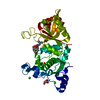
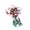
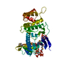
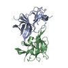
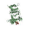
SpikeproteinS1 / S glycoprotein / E2 / Peplomer protein / Spike glycoprotein
Mass: 25520.680 Da / Num. of mol.: 2 / Fragment: receptor-binding domain Source method: isolated from a genetically manipulated source Source: (gene. exp.) Severe acute respiratory syndrome coronavirus 2 Gene: S, 2 / Production host: Spodoptera frugiperda (fall armyworm) / References: UniProt: P0DTC2
#2: Antibody
nanobody
Mass: 13581.988 Da / Num. of mol.: 2 Source method: isolated from a genetically manipulated source Source: (gene. exp.) Lama glama (llama) / Production host: Escherichia coli (E. coli)
In the structure databanks used in Yorodumi, some data are registered as the other names, "COVID-19 virus" and "2019-nCoV". Here are the details of the virus and the list of structure data.
Jan 31, 2019. EMDB accession codes are about to change! (news from PDBe EMDB page)
EMDB accession codes are about to change! (news from PDBe EMDB page)
The allocation of 4 digits for EMDB accession codes will soon come to an end. Whilst these codes will remain in use, new EMDB accession codes will include an additional digit and will expand incrementally as the available range of codes is exhausted. The current 4-digit format prefixed with “EMD-” (i.e. EMD-XXXX) will advance to a 5-digit format (i.e. EMD-XXXXX), and so on. It is currently estimated that the 4-digit codes will be depleted around Spring 2019, at which point the 5-digit format will come into force.
The EM Navigator/Yorodumi systems omit the EMD- prefix.
Related info.:Q: What is EMD? / ID/Accession-code notation in Yorodumi/EM Navigator
Yorodumi is a browser for structure data from EMDB, PDB, SASBDB, etc.
This page is also the successor to EM Navigator detail page, and also detail information page/front-end page for Omokage search.
The word "yorodu" (or yorozu) is an old Japanese word meaning "ten thousand". "mi" (miru) is to see.
Related info.:EMDB / PDB / SASBDB / Comparison of 3 databanks / Yorodumi Search / Aug 31, 2016. New EM Navigator & Yorodumi / Yorodumi Papers / Jmol/JSmol / Function and homology information / Changes in new EM Navigator and Yorodumi
 Movie
Movie Controller


 Open data
 Basic information
 Components
 Keywords
 Function and homology information
 Severe acute respiratory syndrome coronavirus 2
 X-RAY DIFFRACTION /
  Authors
 United States, 3items
  Citation
 Structure visualization
 Downloads & links
 Other downloads

 PDBj
PDBj


 Assembly
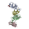


 Spodoptera frugiperda (fall armyworm) / References: UniProt: P0DTC2

 Mass: 35.453 Da / Num. of mol.: 1 / Source method: obtained synthetically / Formula: Cl
 Sample preparation
 Processing