 Movie
Movie Controller


+ Open data

- Basic information
| Entry | Database: PDB / ID: 7kiy | ||||||
|---|---|---|---|---|---|---|---|
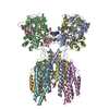
| Title | Plasmodium falciparum RhopH complex in soluble form | ||||||
 Components Components |
| ||||||
 Keywords Keywords | MEMBRANE PROTEIN / Plasmodium falciparum / malaria / RhopH complex | ||||||
| Function / homology | : / RhopH3 C-terminal domain / rhoptry / symbiont-containing vacuole membrane / cytoplasmic vesicle / host cell cytoplasm / host cell plasma membrane / RhopH3 C-terminal domain-containing protein / High molecular weight rhoptry protein 2 Function and homology information Function and homology information | ||||||
| Biological species |  | ||||||
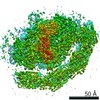
| Method | ELECTRON MICROSCOPY / single particle reconstruction / cryo EM / Resolution: 2.92 Å | ||||||
 Authors Authors | Schureck, M.A. / Darling, J.E. / Merk, A. / Subramaniam, S. / Desai, S.A. | ||||||
| Funding support |  United States, 1items United States, 1items
| ||||||
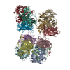
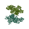
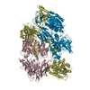
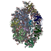
 Citation Citation | Journal: Elife / Year: 2021 Title: Malaria parasites use a soluble RhopH complex for erythrocyte invasion and an integral form for nutrient uptake. Authors: Marc A Schureck / Joseph E Darling / Alan Merk / Jinfeng Shao / Geervani Daggupati / Prakash Srinivasan / Paul Dominic B Olinares / Michael P Rout / Brian T Chait / Kurt Wollenberg / Sriram ...Authors: Marc A Schureck / Joseph E Darling / Alan Merk / Jinfeng Shao / Geervani Daggupati / Prakash Srinivasan / Paul Dominic B Olinares / Michael P Rout / Brian T Chait / Kurt Wollenberg / Sriram Subramaniam / Sanjay A Desai /  Abstract: Malaria parasites use the RhopH complex for erythrocyte invasion and channel-mediated nutrient uptake. As the member proteins are unique to Plasmodium spp., how they interact and traffic through ...Malaria parasites use the RhopH complex for erythrocyte invasion and channel-mediated nutrient uptake. As the member proteins are unique to Plasmodium spp., how they interact and traffic through subcellular sites to serve these essential functions is unknown. We show that RhopH is synthesized as a soluble complex of CLAG3, RhopH2, and RhopH3 with 1:1:1 stoichiometry. After transfer to a new host cell, the complex crosses a vacuolar membrane surrounding the intracellular parasite and becomes integral to the erythrocyte membrane through a PTEX translocon-dependent process. We present a 2.9 Å single-particle cryo-electron microscopy structure of the trafficking complex, revealing that CLAG3 interacts with the other subunits over large surface areas. This soluble complex is tightly assembled with extensive disulfide bonding and predicted transmembrane helices shielded. We propose a large protein complex stabilized for trafficking but poised for host membrane insertion through large-scale rearrangements, paralleling smaller two-state pore-forming proteins in other organisms. | ||||||
| History |
|
- Structure visualization
| Movie |
Movie viewer |
|---|---|
| Structure viewer | Molecule: MolmilJmol/JSmol |
- Downloads & links
-Download
| PDBx/mmCIF format | 7kiy.cif.gz | 481.3 KB | Display | PDBx/mmCIF format |
|---|---|---|---|---|
| PDB format | pdb7kiy.ent.gz | 364.1 KB | Display | PDB format |
| PDBx/mmJSON format | 7kiy.json.gz | Tree view | PDBx/mmJSON format | |
| Others |  Other downloads Other downloads |
-Validation report
| Arichive directory | https://data.pdbj.org/pub/pdb/validation_reports/ki/7kiyftp://data.pdbj.org/pub/pdb/validation_reports/ki/7kiy | HTTPS FTP |
|---|
-Related structure data
| Related structure data | 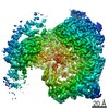 22890MC M: map data used to model this data C: citing same article ( |
|---|---|
| Similar structure data |


-Links
 PDBj
PDBj- Assembly
| Deposited unit | 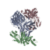
|
|---|---|
| 1 |
|
-Components
| #1: Protein | Mass: 177069.516 Da / Num. of mol.: 1 / Source method: isolated from a natural source Source: (natural) |
|---|---|
| #2: Protein | Mass: 162877.406 Da / Num. of mol.: 1 / Source method: isolated from a natural source Source: (natural) References: UniProt: Q8I060 |
| #3: Protein | Mass: 104981.406 Da / Num. of mol.: 1 / Source method: isolated from a natural source Source: (natural) References: UniProt: A0A024X9S2 |
| Has protein modification | Y |
-Experimental details
-Experiment
| Experiment | Method: ELECTRON MICROSCOPY |
|---|---|
| EM experiment | Aggregation state: PARTICLE / 3D reconstruction method: single particle reconstruction |
- Sample preparation
| Component | Name: RhopH complex / Type: COMPLEX / Entity ID: all / Source: NATURAL | |||||||||||||||
|---|---|---|---|---|---|---|---|---|---|---|---|---|---|---|---|---|
| Molecular weight | Value: 0.434 MDa / Experimental value: YES | |||||||||||||||
| Source (natural) | Organism: | |||||||||||||||
| Buffer solution | pH: 7.5 | |||||||||||||||
| Buffer component |
| |||||||||||||||
| Specimen | Conc.: 0.8 mg/ml / Embedding applied: NO / Shadowing applied: NO / Staining applied: NO / Vitrification applied: YES | |||||||||||||||
| Specimen support | Grid material: COPPER / Grid mesh size: 300 divisions/in. / Grid type: Quantifoil R1.2/1.3 | |||||||||||||||
| Vitrification | Cryogen name: ETHANE / Humidity: 100 % / Chamber temperature: 293.15 K |
- Electron microscopy imaging
| Experimental equipment |  Model: Titan Krios / Image courtesy: FEI Company |
|---|---|
| Microscopy | Model: FEI TITAN KRIOS |
| Electron gun | Electron source:  FIELD EMISSION GUN / Accelerating voltage: 300 kV / Illumination mode: FLOOD BEAM FIELD EMISSION GUN / Accelerating voltage: 300 kV / Illumination mode: FLOOD BEAM |
| Electron lens | Mode: BRIGHT FIELD |
| Image recording | Average exposure time: 0.4 sec. / Electron dose: 70 e/Å2 / Detector mode: COUNTING / Film or detector model: GATAN K2 SUMMIT (4k x 4k) / Num. of grids imaged: 1 / Num. of real images: 1310 |
| Image scans | Movie frames/image: 58 / Used frames/image: 1-58 |
- Processing
| Software |
| ||||||||||||||||||||||||
|---|---|---|---|---|---|---|---|---|---|---|---|---|---|---|---|---|---|---|---|---|---|---|---|---|---|
| EM software |
| ||||||||||||||||||||||||
| Image processing | Details: All cryo-EM image processing was performed in RELION 3.0. | ||||||||||||||||||||||||
| CTF correction | Type: PHASE FLIPPING AND AMPLITUDE CORRECTION | ||||||||||||||||||||||||
| Particle selection | Num. of particles selected: 311390 | ||||||||||||||||||||||||
| 3D reconstruction | Resolution: 2.92 Å / Resolution method: FSC 0.143 CUT-OFF / Num. of particles: 68216 / Num. of class averages: 1 / Symmetry type: POINT | ||||||||||||||||||||||||
| Atomic model building | B value: 97.81 / Protocol: AB INITIO MODEL / Space: REAL | ||||||||||||||||||||||||
| Refinement | Cross valid method: NONE Stereochemistry target values: GeoStd + Monomer Library + CDL v1.2 | ||||||||||||||||||||||||
| Displacement parameters | Biso mean: 97.81 Å2 | ||||||||||||||||||||||||
| Refine LS restraints |
|
