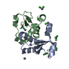
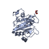
- PDB-7kiq: Crystal structure of the mouse lipin-2 M-Lip domain -
+
Open data
ID or keywords:
Loading...
-
Basic information
Entry
Database: PDB / ID: 7kiq
Title
Crystal structure of the mouse lipin-2 M-Lip domain
Components
Phosphatidate phosphatase LPIN2
Keywords
LIPID BINDING PROTEIN / lipin / M-Lip / dimer
Function / homology
Function and homology information
Synthesis of PE / Triglyceride biosynthesis / phosphatidate phosphatase / Synthesis of PC / Depolymerization of the Nuclear Lamina / phosphatidate phosphatase activity / triglyceride biosynthetic process / fatty acid catabolic process / lipid metabolic process / cellular response to insulin stimulus ...Synthesis of PE / Triglyceride biosynthesis / phosphatidate phosphatase / Synthesis of PC / Depolymerization of the Nuclear Lamina / phosphatidate phosphatase activity / triglyceride biosynthetic process / fatty acid catabolic process / lipid metabolic process / cellular response to insulin stimulus / transcription coactivator activity / endoplasmic reticulum membrane / positive regulation of transcription by RNA polymerase II / nucleus / cytosol Similarity search - Function
In the structure databanks used in Yorodumi, some data are registered as the other names, "COVID-19 virus" and "2019-nCoV". Here are the details of the virus and the list of structure data.
Jan 31, 2019. EMDB accession codes are about to change! (news from PDBe EMDB page)
EMDB accession codes are about to change! (news from PDBe EMDB page)
The allocation of 4 digits for EMDB accession codes will soon come to an end. Whilst these codes will remain in use, new EMDB accession codes will include an additional digit and will expand incrementally as the available range of codes is exhausted. The current 4-digit format prefixed with “EMD-” (i.e. EMD-XXXX) will advance to a 5-digit format (i.e. EMD-XXXXX), and so on. It is currently estimated that the 4-digit codes will be depleted around Spring 2019, at which point the 5-digit format will come into force.
The EM Navigator/Yorodumi systems omit the EMD- prefix.
Related info.:Q: What is EMD? / ID/Accession-code notation in Yorodumi/EM Navigator
Yorodumi is a browser for structure data from EMDB, PDB, SASBDB, etc.
This page is also the successor to EM Navigator detail page, and also detail information page/front-end page for Omokage search.
The word "yorodu" (or yorozu) is an old Japanese word meaning "ten thousand". "mi" (miru) is to see.
Related info.:EMDB / PDB / SASBDB / Comparison of 3 databanks / Yorodumi Search / Aug 31, 2016. New EM Navigator & Yorodumi / Yorodumi Papers / Jmol/JSmol / Function and homology information / Changes in new EM Navigator and Yorodumi
 Movie
Movie Controller


 Open data
 Basic information
 Components
 Keywords
 Function and homology information
 X-RAY DIFFRACTION /
  Authors
 United States, 2items
  Citation
 Structure visualization
 Downloads & links
 Other downloads
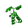


 PDBj
PDBj Assembly


 Mass: 40.078 Da / Num. of mol.: 10 / Source method: obtained synthetically / Formula: Ca
 Mass: 18.015 Da / Num. of mol.: 254 / Source method: isolated from a natural source / Formula: H2O
 Sample preparation
 Processing