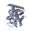
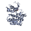
[English] 日本語
Yorodumi
- PDB-7kht: The acyl chains of phosphoinositide PIP3 alter the structure and ... -

+
Open data


ID or keywords:

Loading...

-
Basic information

Entry
Database: PDB / ID: 7kht
TitleThe acyl chains of phosphoinositide PIP3 alter the structure and function of nuclear receptor Steroidogenic Factor-1 (SF-1)
Components
  • Peroxisome proliferator-activated receptor gamma coactivator 1-alpha peptide
  • Steroidogenic factor 1
KeywordsLIPID BINDING PROTEIN / PI(3 / 4 / 5)P3 acyl chains / acyl chain length / acyl chain induced conformational change / nuclear PIP2 / nuclear PIP3 / nuclear receptor
Function / homology
Function and homology information


primary sex determination / response to gonadotropin-releasing hormone / Sertoli cell differentiation / negative regulation of female gonad development / positive regulation of mitochondrial DNA metabolic process / positive regulation of muscle tissue development / sex determination / regulation of steroid biosynthetic process / positive regulation of male gonad development / Transcriptional regulation of testis differentiation ...primary sex determination / response to gonadotropin-releasing hormone / Sertoli cell differentiation / negative regulation of female gonad development / positive regulation of mitochondrial DNA metabolic process / positive regulation of muscle tissue development / sex determination / regulation of steroid biosynthetic process / positive regulation of male gonad development / Transcriptional regulation of testis differentiation / luteinization / calcineurin-mediated signaling / positive regulation of cellular respiration / Transcriptional regulation of pluripotent stem cells / positive regulation of fatty acid oxidation / tissue development / Leydig cell differentiation / : / male sex determination / maintenance of protein location in nucleus / hormone metabolic process / : / lncRNA binding / cellular respiration / response to muscle activity / Activation of PPARGC1A (PGC-1alpha) by phosphorylation / temperature homeostasis / adrenal gland development / female gonad development / positive regulation of ATP biosynthetic process / response to starvation / intracellular glucose homeostasis / fatty acid oxidation / response to dietary excess / adipose tissue development / FOXO-mediated transcription of oxidative stress, metabolic and neuronal genes / energy homeostasis / brown fat cell differentiation / positive regulation of gluconeogenesis / digestion / respiratory electron transport chain / hormone-mediated signaling pathway / mitochondrion organization / SUMOylation of transcription cofactors / nuclear receptor coactivator activity / RNA splicing / gluconeogenesis / transcription coregulator binding / transcription initiation at RNA polymerase II promoter / nuclear receptor binding / transcription coregulator activity / negative regulation of smooth muscle cell proliferation / RNA polymerase II transcription regulatory region sequence-specific DNA binding / phospholipid binding / circadian regulation of gene expression / Heme signaling / SUMOylation of intracellular receptors / Transcriptional activation of mitochondrial biogenesis / regulation of circadian rhythm / PPARA activates gene expression / PML body / chromatin DNA binding / mRNA processing / Transcriptional regulation of white adipocyte differentiation / Nuclear Receptor transcription pathway / RNA polymerase II transcription regulator complex / male gonad development / Regulation of RUNX2 expression and activity / nuclear receptor activity / positive regulation of DNA-binding transcription factor activity / Circadian Clock / sequence-specific double-stranded DNA binding / cellular response to oxidative stress / positive regulation of cold-induced thermogenesis / protein-containing complex assembly / neuron apoptotic process / DNA-binding transcription factor binding / negative regulation of neuron apoptotic process / RNA polymerase II-specific DNA-binding transcription factor binding / sequence-specific DNA binding / transcription by RNA polymerase II / transcription coactivator activity / protein stabilization / DNA-binding transcription factor activity, RNA polymerase II-specific / RNA polymerase II cis-regulatory region sequence-specific DNA binding / ubiquitin protein ligase binding / chromatin binding / chromatin / positive regulation of gene expression / regulation of DNA-templated transcription / regulation of transcription by RNA polymerase II / positive regulation of DNA-templated transcription / enzyme binding / positive regulation of transcription by RNA polymerase II / DNA binding / RNA binding / zinc ion binding / nucleoplasm / nucleus / cytosol
Similarity search - Function
PGC-1alpha, RNA recognition motif / PGC-1 / Nuclear hormone receptor family 5 / RNA recognition motif / RNA recognition motif / Eukaryotic RNA Recognition Motif (RRM) profile. / RNA recognition motif domain / RNA-binding domain superfamily / Nuclear hormone receptor / Nuclear hormones receptors DNA-binding region signature. ...PGC-1alpha, RNA recognition motif / PGC-1 / Nuclear hormone receptor family 5 / RNA recognition motif / RNA recognition motif / Eukaryotic RNA Recognition Motif (RRM) profile. / RNA recognition motif domain / RNA-binding domain superfamily / Nuclear hormone receptor / Nuclear hormones receptors DNA-binding region signature. / Zinc finger, nuclear hormone receptor-type / Zinc finger, C4 type (two domains) / Nuclear hormone receptors DNA-binding domain profile. / c4 zinc finger in nuclear hormone receptors / Nuclear hormone receptor, ligand-binding domain / Nuclear hormone receptor-like domain superfamily / Ligand-binding domain of nuclear hormone receptor / Nuclear receptor (NR) ligand-binding (LBD) domain profile. / Ligand binding domain of hormone receptors / Zinc finger, NHR/GATA-type / Nucleotide-binding alpha-beta plait domain superfamily
Similarity search - Domain/homology
Chem-WES / Steroidogenic factor 1 / Peroxisome proliferator-activated receptor gamma coactivator 1-alpha
Similarity search - Component
Biological speciesHomo sapiens (human)
MethodX-RAY DIFFRACTION / SYNCHROTRON / MOLECULAR REPLACEMENT / Resolution: 2.504 Å
AuthorsBlind, R.D.
Funding support United States, 4items
OrganizationGrant numberCountry
American Cancer SocietyRSG-17-063-01 United States
National Institutes of Health/National Institute of General Medical Sciences (NIH/NIGMS)GM138873 United States
National Institutes of Health/National Cancer Institute (NIH/NCI)CA243036 United States
National Institutes of Health/National Institute of Diabetes and Digestive and Kidney Disease (NIH/NIDDK) United States
CitationJournal: J.Lipid Res. / Year: 2021
Title: The acyl chains of phosphoinositide PIP3 alter the structure and function of nuclear receptor steroidogenic factor-1.
Authors: Bryant, J.M. / Malabanan, M.M. / Vanderloop, B.H. / Nichols, C.M. / Haratipour, Z. / Poon, K.T. / Sherrod, S.D. / McLean, J.A. / Blind, R.D.
History
DepositionOct 22, 2020Deposition site: RCSB / Processing site: RCSB
Revision 1.0May 19, 2021Provider: repository / Type: Initial release
Revision 1.1Jun 9, 2021Group: Database references / Category: citation / Item: _citation.journal_volume / _citation.title
Revision 1.2Oct 18, 2023Group: Data collection / Database references / Refinement description
Category: chem_comp_atom / chem_comp_bond ...chem_comp_atom / chem_comp_bond / database_2 / pdbx_initial_refinement_model
Item: _database_2.pdbx_DOI / _database_2.pdbx_database_accession

-
Structure visualization

Structure viewerMolecule:
MolmilJmol/JSmol

Downloads & links

-
Assembly

Deposited unit
A: Steroidogenic factor 1
B: Peroxisome proliferator-activated receptor gamma coactivator 1-alpha peptide
hetero molecules


Theoretical massNumber of molelcules
Total (without water)30,5303
Polymers29,4272
Non-polymers1,1031
Water59433
1


  • Idetical with deposited unit
  • defined by author&software
  • Evidence: assay for oligomerization, PIP3 alters anion exchange mobility due to additional charges on headgroup. Peptide was ordered in structure.
TypeNameSymmetry operationNumber
identity operation1_555x,y,z1
Buried area930 Å2
ΔGint-11 kcal/mol
Surface area12790 Å2
MethodPISA
Unit cell
Length a, b, c (Å)65.349, 65.349, 139.676
Angle α, β, γ (deg.)90.00, 90.00, 90.00
Int Tables number92
Space group name H-MP41212

-
Components

#1: Protein Steroidogenic factor 1 / / hSF-1 / Adrenal 4-binding protein / Fushi tarazu factor homolog 1 / Nuclear receptor subfamily 5 ...hSF-1 / Adrenal 4-binding protein / Fushi tarazu factor homolog 1 / Nuclear receptor subfamily 5 group A member 1 / Steroid hormone receptor Ad4BP


Mass: 27903.406 Da / Num. of mol.: 1 / Mutation: C247S, C412S
Source method: isolated from a genetically manipulated source
Source: (gene. exp.) Homo sapiens (human) / Gene: NR5A1, AD4BP, FTZF1, SF1 / Production host: Escherichia coli (E. coli) / References: UniProt: Q13285
#2: Protein/peptide Peroxisome proliferator-activated receptor gamma coactivator 1-alpha peptide / PPARGC-1-alpha / Ligand effect modulator 6


Mass: 1523.854 Da / Num. of mol.: 1 / Source method: obtained synthetically / Source: (synth.) Homo sapiens (human) / References: UniProt: Q9UBK2
#3: Chemical ChemComp-WES / (2S)-3-{[(S)-{[(1S,2S,3R,4S,5S,6S)-2,6-dihydroxy-3,4,5-tris(phosphonooxy)cyclohexyl]oxy}(hydroxy)phosphoryl]oxy}propane-1,2-diyl (9E,9'E)di-octadec-9-enoate


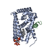
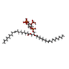
Mass: 1103.046 Da / Num. of mol.: 1 / Source method: obtained synthetically / Formula: C45H86O22P4
#4: Water ChemComp-HOH / water / Water


Mass: 18.015 Da / Num. of mol.: 33 / Source method: isolated from a natural source / Formula: H2O
Has ligand of interestY

-
Experimental details

-
Experiment

ExperimentMethod: X-RAY DIFFRACTION / Number of used crystals: 1

-
Sample preparation

CrystalDensity Matthews: 2.65 Å3/Da / Density % sol: 53.65 %
Crystal growTemperature: 293 K / Method: vapor diffusion, sitting drop / Details: 20% PEG 3350 and 0.2 M magnesium formate

-
Data collection

DiffractionMean temperature: 100 K / Serial crystal experiment: N
Diffraction sourceSource: SYNCHROTRON / Site: APS / Beamline: 21-ID-F / Wavelength: 0.97857 Å
DetectorType: RAYONIX MX-300 / Detector: CCD / Date: Jun 20, 2018
RadiationProtocol: SINGLE WAVELENGTH / Monochromatic (M) / Laue (L): M / Scattering type: x-ray
Radiation wavelengthWavelength: 0.97857 Å / Relative weight: 1
ReflectionResolution: 2.5→38.54 Å / Num. obs: 21999 / % possible obs: 98.38 % / Redundancy: 2 % / Biso Wilson estimate: 70.45 Å2 / CC1/2: 1 / CC star: 1 / Net I/σ(I): 21.8
Reflection shellResolution: 2.504→2.594 Å / Redundancy: 2 % / Mean I/σ(I) obs: 2.6 / Num. unique obs: 1053 / CC1/2: 0.912 / CC star: 0.977 / % possible all: 98.78

-
Processing

Software
NameVersionClassification
PHENIX(1.14_3260: ???)refinement
PDB_EXTRACT3.25data extraction
HKL-2000data reduction
HKL-2000data scaling
PHASERphasing
RefinementMethod to determine structure: MOLECULAR REPLACEMENT
Starting model: 4QJR
Resolution: 2.504→38.537 Å / SU ML: 0.37 / Cross valid method: THROUGHOUT / σ(F): 0.23 / Phase error: 32.41 / Stereochemistry target values: ML
RfactorNum. reflection% reflection
Rfree0.2488 911 4.65 %
Rwork0.2187 --
obs0.2202 19587 98.51 %
Solvent computationShrinkage radii: 0.9 Å / VDW probe radii: 1.11 Å / Solvent model: FLAT BULK SOLVENT MODEL
Refinement stepCycle: LAST / Resolution: 2.504→38.537 Å
ProteinNucleic acidLigandSolventTotal
Num. atoms1945 0 66 33 2044
Refine LS restraints
Refine-IDTypeDev idealNumber
X-RAY DIFFRACTIONf_bond_d0.012043
X-RAY DIFFRACTIONf_angle_d1.2972767
X-RAY DIFFRACTIONf_dihedral_angle_d20.672816
X-RAY DIFFRACTIONf_chiral_restr0.077335
X-RAY DIFFRACTIONf_plane_restr0.007342
LS refinement shell
Resolution (Å)Rfactor RfreeNum. reflection RfreeRfactor RworkNum. reflection RworkRefine-ID% reflection obs (%)
2.5044-2.63640.39011180.3492683X-RAY DIFFRACTION99
2.6364-2.80150.34221450.34422700X-RAY DIFFRACTION99
2.8015-3.01770.33681530.32592621X-RAY DIFFRACTION99
3.0177-3.32130.2692850.29352720X-RAY DIFFRACTION98
3.3213-3.80150.31981320.23392633X-RAY DIFFRACTION97
3.8015-4.78810.19491250.18432645X-RAY DIFFRACTION98
4.7881-38.5370.21951530.1762674X-RAY DIFFRACTION99
Refinement TLS params.

Method: refined / Refine-ID: X-RAY DIFFRACTION

IDL112)L122)L132)L222)L232)L332)S11 (Å °)S12 (Å °)S13 (Å °)S21 (Å °)S22 (Å °)S23 (Å °)S31 (Å °)S32 (Å °)S33 (Å °)T112)T122)T132)T222)T232)T332)Origin x (Å)Origin y (Å)Origin z (Å)
17.8918-0.2708-1.06236.28251.47766.35350.18680.00582.55970.89420.32310.8223-1.0608-0.0022-0.53681.3058-0.0170.19740.7380.1141.31335.5001-5.01891.3179
23.7461.46440.27764.39381.79975.42590.5610.94870.6829-1.04670.19790.3659-0.58090.1829-0.45521.20920.0570.01751.1580.32840.977320.389-16.7878-19.0146
35.81880.01351.38.36745.32225.00880.34050.40291.1477-0.71780.6509-1.0328-1.25590.8187-1.04320.9981-0.18250.10280.81880.10660.952323.1721-14.2074-8.3474
42.7717-2.402-1.23296.8342.45754.3302-0.0140.25110.2375-0.20580.0418-0.17980.05020.10620.03140.6534-0.06350.03190.71980.13140.57516.5759-24.6717-7.6782
56.9037-2.5299-1.40633.68810.24274.6004-0.0957-0.29170.01890.13970.1289-0.07990.1337-0.220.03260.6945-0.05760.02010.6363-0.04580.551810.9224-24.0938-0.6914
67.6346-5.0232.11643.3778-0.70586.7625-1.4859-2.85542.0214-0.4760.84-1.8353-0.2377-0.3658-0.00551.1757-0.0341-0.31241.3507-0.27561.403327.4631-12.129812.2857
73.6778-1.9104-2.16145.41822.43695.44120.5183-0.52111.07450.9694-0.25530.2632-0.1840.2673-0.38310.9623-0.06850.05950.7851-0.14780.8227.6388-11.833810.4528
86.9558-3.3564-2.64112.6991.61774.4554-0.5047-1.4835-0.55030.28340.40280.2294-0.140.79180.03920.6372-0.00750.02980.63470.02670.600519.7164-27.93132.2224
92.6163-3.0871-3.02523.90513.81343.7819-0.4349-0.8762-0.1894-0.59460.8907-1.5882-0.33222.7421-0.21770.904-0.04480.10711.2366-0.03521.15233.7464-25.55-8.8927
107.9568-5.21920.4043.46810.07362.44370.11761.47491.1143-0.4723-1.1554-2.45380.15960.38040.57851.4384-0.35310.32761.1834-0.11851.782132.6873-8.906-7.401
Refinement TLS group
IDRefine-IDRefine TLS-IDSelection details
1X-RAY DIFFRACTION1chain 'A' and (resid 221 through 232 )
2X-RAY DIFFRACTION2chain 'A' and (resid 233 through 259 )
3X-RAY DIFFRACTION3chain 'A' and (resid 260 through 290 )
4X-RAY DIFFRACTION4chain 'A' and (resid 291 through 341 )
5X-RAY DIFFRACTION5chain 'A' and (resid 342 through 377 )
6X-RAY DIFFRACTION6chain 'A' and (resid 378 through 386 )
7X-RAY DIFFRACTION7chain 'A' and (resid 387 through 414 )
8X-RAY DIFFRACTION8chain 'A' and (resid 415 through 442 )
9X-RAY DIFFRACTION9chain 'A' and (resid 443 through 458 )
10X-RAY DIFFRACTION10chain 'B' and (resid 141 through 152 )

+
About Yorodumi

-
News

-
Feb 9, 2022. New format data for meta-information of EMDB entries

New format data for meta-information of EMDB entries

  • Version 3 of the EMDB header file is now the official format.
  • The previous official version 1.9 will be removed from the archive.

Related info.:EMDB header

External links:wwPDB to switch to version 3 of the EMDB data model

-
Aug 12, 2020. Covid-19 info

Covid-19 info

URL: https://pdbj.org/emnavi/covid19.php

New page: Covid-19 featured information page in EM Navigator.

Related info.:Covid-19 info / Mar 5, 2020. Novel coronavirus structure data

+
Mar 5, 2020. Novel coronavirus structure data

Novel coronavirus structure data

Related info.:Yorodumi Speices / Aug 12, 2020. Covid-19 info

External links:COVID-19 featured content - PDBj / Molecule of the Month (242):Coronavirus Proteases

+
Jan 31, 2019. EMDB accession codes are about to change! (news from PDBe EMDB page)

EMDB accession codes are about to change! (news from PDBe EMDB page)

  • The allocation of 4 digits for EMDB accession codes will soon come to an end. Whilst these codes will remain in use, new EMDB accession codes will include an additional digit and will expand incrementally as the available range of codes is exhausted. The current 4-digit format prefixed with “EMD-” (i.e. EMD-XXXX) will advance to a 5-digit format (i.e. EMD-XXXXX), and so on. It is currently estimated that the 4-digit codes will be depleted around Spring 2019, at which point the 5-digit format will come into force.
  • The EM Navigator/Yorodumi systems omit the EMD- prefix.

Related info.:Q: What is EMD? / ID/Accession-code notation in Yorodumi/EM Navigator

External links:EMDB Accession Codes are Changing Soon! / Contact to PDBj

+
Jul 12, 2017. Major update of PDB

Major update of PDB

  • wwPDB released updated PDB data conforming to the new PDBx/mmCIF dictionary.
  • This is a major update changing the version number from 4 to 5, and with Remediation, in which all the entries are updated.
  • In this update, many items about electron microscopy experimental information are reorganized (e.g. em_software).
  • Now, EM Navigator and Yorodumi are based on the updated data.

External links:wwPDB Remediation / Enriched Model Files Conforming to OneDep Data Standards Now Available in the PDB FTP Archive

-
Yorodumi

Thousand views of thousand structures

  • Yorodumi is a browser for structure data from EMDB, PDB, SASBDB, etc.
  • This page is also the successor to EM Navigator detail page, and also detail information page/front-end page for Omokage search.
  • The word "yorodu" (or yorozu) is an old Japanese word meaning "ten thousand". "mi" (miru) is to see.

Related info.:EMDB / PDB / SASBDB / Comparison of 3 databanks / Yorodumi Search / Aug 31, 2016. New EM Navigator & Yorodumi / Yorodumi Papers / Jmol/JSmol / Function and homology information / Changes in new EM Navigator and Yorodumi

Read more