 Movie
Movie Controller


+ Open data

- Basic information
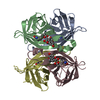
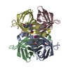
| Entry | Database: PDB / ID: 7kby | ||||||
|---|---|---|---|---|---|---|---|
| Title | Artificial Metalloproteins with Dinuclear Iron Centers | ||||||
 Components Components | Streptavidin | ||||||
 Keywords Keywords | METAL BINDING PROTEIN / Biotin binding Artificial Metalloprotein | ||||||
| Function / homology |  Function and homology information Function and homology information | ||||||
| Biological species |  Streptomyces avidinii (bacteria) Streptomyces avidinii (bacteria) | ||||||
| Method |  X-RAY DIFFRACTION / SYNCHROTRON / MOLECULAR REPLACEMENT / Resolution: 1.7 Å X-RAY DIFFRACTION / SYNCHROTRON / MOLECULAR REPLACEMENT / Resolution: 1.7 Å | ||||||
 Authors Authors | Miller, K.R. / Follmer, A.H. / Jasniewski, A.J. / Sabuncu, S. / Biswas, S. / Albert, T. / Hendrich, M.P. / Moenne-Loccoz, P. / Borovik, A.S. | ||||||
| Funding support |  United States, 1items United States, 1items
| ||||||
 Citation Citation | Journal: J.Am.Chem.Soc. / Year: 2021 Title: Artificial Metalloproteins with Dinuclear Iron-Hydroxido Centers. Authors: Miller, K.R. / Biswas, S. / Jasniewski, A. / Follmer, A.H. / Biswas, A. / Albert, T. / Sabuncu, S. / Bominaar, E.L. / Hendrich, M.P. / Moenne-Loccoz, P. / Borovik, A.S. | ||||||
| History |
|
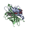
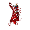
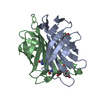
- Structure visualization
| Structure viewer | Molecule: MolmilJmol/JSmol |
|---|
- Downloads & links
-Download
| PDBx/mmCIF format | 7kby.cif.gz | 44 KB | Display | PDBx/mmCIF format |
|---|---|---|---|---|
| PDB format | pdb7kby.ent.gz | 27.6 KB | Display | PDB format |
| PDBx/mmJSON format | 7kby.json.gz | Tree view | PDBx/mmJSON format | |
| Others |  Other downloads Other downloads |
-Validation report
| Arichive directory | https://data.pdbj.org/pub/pdb/validation_reports/kb/7kbyftp://data.pdbj.org/pub/pdb/validation_reports/kb/7kby | HTTPS FTP |
|---|
-Related structure data
| Related structure data | 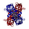 6vo9C 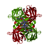 6vobC 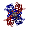 6vozC 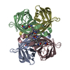 6vp1C 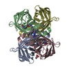 6vp2C  6vp3C  7kbzC  7knlC  2qcbS S: Starting model for refinement C: citing same article ( |
|---|---|
| Similar structure data |


-Links
 PDBj
PDBj


- Assembly
| Deposited unit | 
| |||||||||||||||
|---|---|---|---|---|---|---|---|---|---|---|---|---|---|---|---|---|
| 1 | 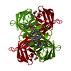
| |||||||||||||||
| Unit cell |
| |||||||||||||||
| Components on special symmetry positions |
|
-Components
| #1: Protein | Mass: 16561.932 Da / Num. of mol.: 1 / Mutation: K121A, L124Y Source method: isolated from a genetically manipulated source Source: (gene. exp.) Streptomyces avidinii (bacteria) / Production host: |
|---|---|
| #2: Chemical | ChemComp-KM3 / {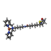  Mass: 552.513 Da / Num. of mol.: 1 / Source method: obtained synthetically / Formula: C26H36FeN6O2S / Feature type: SUBJECT OF INVESTIGATION Mass: 552.513 Da / Num. of mol.: 1 / Source method: obtained synthetically / Formula: C26H36FeN6O2S / Feature type: SUBJECT OF INVESTIGATION |
| #3: Chemical | ChemComp-CYN /   Mass: 26.017 Da / Num. of mol.: 1 / Source method: obtained synthetically / Formula: CN Mass: 26.017 Da / Num. of mol.: 1 / Source method: obtained synthetically / Formula: CN |
| #4: Chemical | ChemComp-ACT /   Mass: 59.044 Da / Num. of mol.: 1 / Source method: obtained synthetically / Formula: C2H3O2 Mass: 59.044 Da / Num. of mol.: 1 / Source method: obtained synthetically / Formula: C2H3O2 |
| #5: Water | ChemComp-HOH /  Mass: 18.015 Da / Num. of mol.: 70 / Source method: isolated from a natural source / Formula: H2O Mass: 18.015 Da / Num. of mol.: 70 / Source method: isolated from a natural source / Formula: H2O |
| Has ligand of interest | Y |
-Experimental details
-Experiment
| Experiment | Method: X-RAY DIFFRACTION / Number of used crystals: 1 |
|---|
- Sample preparation
| Crystal | Density Matthews: 2.29 Å3/Da / Density % sol: 46.37 % |
|---|---|
| Crystal grow | Temperature: 295 K / Method: vapor diffusion, sitting drop / pH: 4 Details: 26 mg/mL 2.0 M ammonium sulfate, 0.1 M sodium acetate pH 4 |
-Data collection
| Diffraction | Mean temperature: 100 K / Serial crystal experiment: N | ||||||||||||||||||||||||||||||
|---|---|---|---|---|---|---|---|---|---|---|---|---|---|---|---|---|---|---|---|---|---|---|---|---|---|---|---|---|---|---|---|
| Diffraction source | Source: SYNCHROTRON / Site: ALS / Beamline: 5.0.2 / Wavelength: 1 Å | ||||||||||||||||||||||||||||||
| Detector | Type: DECTRIS PILATUS3 6M / Detector: PIXEL / Date: Mar 16, 2019 | ||||||||||||||||||||||||||||||
| Radiation | Protocol: SINGLE WAVELENGTH / Monochromatic (M) / Laue (L): M / Scattering type: x-ray | ||||||||||||||||||||||||||||||
| Radiation wavelength | Wavelength: 1 Å / Relative weight: 1 | ||||||||||||||||||||||||||||||
| Reflection | Resolution: 1.7→45.86 Å / Num. obs: 17537 / % possible obs: 100 % / Redundancy: 11.7 % / CC1/2: 0.997 / Rmerge(I) obs: 0.21 / Rpim(I) all: 0.063 / Rrim(I) all: 0.22 / Net I/σ(I): 9.1 / Num. measured all: 205175 / Scaling rejects: 43 | ||||||||||||||||||||||||||||||
| Reflection shell | Diffraction-ID: 1
|
- Processing
| Software |
| ||||||||||||||||||||||||||||||||||||||||||||||||||||||||||||
|---|---|---|---|---|---|---|---|---|---|---|---|---|---|---|---|---|---|---|---|---|---|---|---|---|---|---|---|---|---|---|---|---|---|---|---|---|---|---|---|---|---|---|---|---|---|---|---|---|---|---|---|---|---|---|---|---|---|---|---|---|---|
| Refinement | Method to determine structure: MOLECULAR REPLACEMENT Starting model: 2QCB Resolution: 1.7→41.95 Å / Cor.coef. Fo:Fc: 0.959 / Cor.coef. Fo:Fc free: 0.943 / Cross valid method: THROUGHOUT / σ(F): 0 / ESU R: 0.097 / ESU R Free: 0.098 / Stereochemistry target values: MAXIMUM LIKELIHOOD Details: HYDROGENS HAVE BEEN ADDED IN THE RIDING POSITIONS U VALUES : REFINED INDIVIDUALLY
| ||||||||||||||||||||||||||||||||||||||||||||||||||||||||||||
| Solvent computation | Ion probe radii: 0.8 Å / Shrinkage radii: 0.8 Å / VDW probe radii: 1.2 Å / Solvent model: MASK | ||||||||||||||||||||||||||||||||||||||||||||||||||||||||||||
| Displacement parameters | Biso max: 79.81 Å2 / Biso mean: 20.95 Å2 / Biso min: 10.97 Å2
| ||||||||||||||||||||||||||||||||||||||||||||||||||||||||||||
| Refinement step | Cycle: final / Resolution: 1.7→41.95 Å
| ||||||||||||||||||||||||||||||||||||||||||||||||||||||||||||
| Refine LS restraints |
| ||||||||||||||||||||||||||||||||||||||||||||||||||||||||||||
| LS refinement shell | Resolution: 1.7→1.744 Å / Rfactor Rfree error: 0 / Total num. of bins used: 20
|
