[English] 日本語
Yorodumi
- PDB-6gmi: Genetic Engineering of an Artificial Metalloenzyme for Transfer H... -

+
Open data


ID or keywords:

Loading...

-
Basic information

Entry
Database: PDB / ID: 6gmi
TitleGenetic Engineering of an Artificial Metalloenzyme for Transfer Hydrogenation of a Self-Immolative Substrate in E. coli's Periplasm.
ComponentsStreptavidin
Keywordsbiotin-binding protein / Artificial Transfer Hydrogenase / Beta Barrel / Streptavidin
Function / homology
Function and homology information


biotin binding / extracellular region
Similarity search - Function
Avidin-like / Avidin-like, conserved site / Avidin-like domain signature. / Avidin / Avidin/streptavidin / Avidin-like superfamily / Avidin family / Avidin-like domain profile. / Lipocalin / Beta Barrel / Mainly Beta
Similarity search - Domain/homology
Chem-4IR / IRIDIUM (III) ION / Streptavidin
Similarity search - Component
Biological speciesStreptomyces avidinii (bacteria)
MethodX-RAY DIFFRACTION / SYNCHROTRON / MOLECULAR REPLACEMENT / Resolution: 1.6 Å
AuthorsRebelein, J.G.
Funding support Switzerland, Germany, 4items
OrganizationGrant numberCountry
Swiss National Science Foundation200020_162348 Switzerland
European Research CouncilDrEAM
Swiss National Science FoundationNCCR MSE Switzerland
European Molecular Biology OrganizationALTF 194-2017 Germany
CitationJournal: J. Am. Chem. Soc. / Year: 2018
Title: Genetic Engineering of an Artificial Metalloenzyme for Transfer Hydrogenation of a Self-Immolative Substrate in Escherichia coli's Periplasm.
Authors: Zhao, J. / Rebelein, J.G. / Mallin, H. / Trindler, C. / Pellizzoni, M.M. / Ward, T.R.
History
DepositionMay 26, 2018Deposition site: PDBE / Processing site: PDBE
Revision 1.0Oct 10, 2018Provider: repository / Type: Initial release
Revision 1.1Oct 31, 2018Group: Data collection / Database references / Structure summary
Category: citation / entity
Item: _citation.journal_volume / _citation.page_first ..._citation.journal_volume / _citation.page_first / _citation.page_last / _entity.formula_weight
Revision 1.2Jan 17, 2024Group: Data collection / Database references / Refinement description
Category: chem_comp_atom / chem_comp_bond ...chem_comp_atom / chem_comp_bond / database_2 / pdbx_initial_refinement_model
Item: _database_2.pdbx_DOI / _database_2.pdbx_database_accession

-
Structure visualization

Structure viewerMolecule:
MolmilJmol/JSmol

Downloads & links

-
Assembly

Deposited unit
A: Streptavidin
hetero molecules


Theoretical massNumber of molelcules
Total (without water)20,2016
Polymers18,6241
Non-polymers1,5765
Water61334
1
A: Streptavidin
hetero molecules

A: Streptavidin
hetero molecules

A: Streptavidin
hetero molecules

A: Streptavidin
hetero molecules


Theoretical massNumber of molelcules
Total (without water)80,80224
Polymers74,4974
Non-polymers6,30520
Water724
TypeNameSymmetry operationNumber
identity operation1_555x,y,z1
crystal symmetry operation8_665-y+1,-x+1,-z1
crystal symmetry operation10_665-x+1,-y+1,z1
crystal symmetry operation15_555y,x,-z1
Buried area11680 Å2
ΔGint-161 kcal/mol
Surface area17630 Å2
MethodPISA
Unit cell
Length a, b, c (Å)57.507, 57.507, 184.130
Angle α, β, γ (deg.)90.00, 90.00, 90.00
Int Tables number98
Space group name H-MI4122
Components on special symmetry positions
IDModelComponents
11A-302-

HOH

21A-307-

HOH

-
Components

#1: Protein Streptavidin


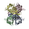
Mass: 18624.266 Da / Num. of mol.: 1 / Mutation: S112V, E116SPLSEALTKANSPAEAYKASRGAGA, K121A
Source method: isolated from a genetically manipulated source
Details: T7-Tag on the N-Terminus of Streptavidin, Insertion of SPLSEALTKANSPAEAYKASRGAGA for E116
Source: (gene. exp.) Streptomyces avidinii (bacteria) / Production host: Escherichia coli (E. coli) / References: UniProt: P22629
#2: Chemical ChemComp-4IR / {N-(4-{[2-(amino-kappaN)ethyl]sulfamoyl-kappaN}phenyl)-5-[(3aS,4S,6aR)-2-oxohexahydro-1H-thieno[3,4-d]imidazol-4-yl]pentanamide}(chloro)[(1,2,3,4,5-eta)-1,2,3,4,5-pentamethylcyclopentadienyl]iridium(III) / N-(4-{[(2-AMINOETHYL)AMINO]SULFONYL}PHENYL)-5-[(3AS,4S,6AR)-2-OXOHEXAHYDRO-1H-THIENO[3,4-D]IMIDAZOL-4-YL]PENTANAMIDE-(1,2,3,4,5,6-ETA)-PENTAMETHYLCYCLOHEXYL-CHLORO-IRIDIUM(III)


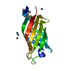
Mass: 807.488 Da / Num. of mol.: 1 / Source method: obtained synthetically / Formula: C28H45ClIrN5O4S2
#3: Chemical
ChemComp-IR3 / IRIDIUM (III) ION


Mass: 192.217 Da / Num. of mol.: 4 / Source method: obtained synthetically / Formula: Ir
#4: Water ChemComp-HOH / water


Mass: 18.015 Da / Num. of mol.: 34 / Source method: isolated from a natural source / Formula: H2O

-
Experimental details

-
Experiment

ExperimentMethod: X-RAY DIFFRACTION / Number of used crystals: 1

-
Sample preparation

CrystalDensity Matthews: 2.04 Å3/Da / Density % sol: 39.81 %
Crystal growTemperature: 293 K / Method: vapor diffusion, sitting drop / pH: 9
Details: 0.1 M SPG 9 pH (Buffer) 25 %w/v PEG 1500 (Precipitant)

-
Data collection

DiffractionMean temperature: 100 K
Diffraction sourceSource: SYNCHROTRON / Site: SLS / Beamline: X06DA / Wavelength: 1 Å
DetectorType: DECTRIS EIGER X 16M / Detector: PIXEL / Date: May 18, 2018
RadiationProtocol: SINGLE WAVELENGTH / Monochromatic (M) / Laue (L): M / Scattering type: x-ray
Radiation wavelengthWavelength: 1 Å / Relative weight: 1
ReflectionResolution: 1.6→46.07 Å / Num. obs: 20991 / % possible obs: 100 % / Redundancy: 25.4 % / Rmerge(I) obs: 0.117 / Net I/σ(I): 15.6
Reflection shellResolution: 1.6→1.63 Å / Rmerge(I) obs: 4.33

-
Processing

Software
NameVersionClassification
REFMAC5.8.0222refinement
XDSdata reduction
Aimlessdata scaling
PHASERphasing
RefinementMethod to determine structure: MOLECULAR REPLACEMENT
Starting model: 2qcb
Resolution: 1.6→46.07 Å / Cor.coef. Fo:Fc: 0.968 / Cor.coef. Fo:Fc free: 0.97 / SU B: 1.7 / SU ML: 0.057 / Cross valid method: THROUGHOUT / ESU R: 0.076 / ESU R Free: 0.073 / Details: HYDROGENS HAVE BEEN ADDED IN THE RIDING POSITIONS
RfactorNum. reflection% reflectionSelection details
Rfree0.1994 1065 5.1 %RANDOM
Rwork0.18661 ---
obs0.18728 19899 99.98 %-
Solvent computationIon probe radii: 0.8 Å / Shrinkage radii: 0.8 Å / VDW probe radii: 1.2 Å
Displacement parametersBiso mean: 30.467 Å2
Baniso -1Baniso -2Baniso -3
1--0.37 Å20 Å20 Å2
2---0.37 Å20 Å2
3---0.75 Å2
Refinement stepCycle: 1 / Resolution: 1.6→46.07 Å
ProteinNucleic acidLigandSolventTotal
Num. atoms896 0 45 34 975
Refine LS restraints
Refine-IDTypeDev idealDev ideal targetNumber
X-RAY DIFFRACTIONr_bond_refined_d0.0150.014988
X-RAY DIFFRACTIONr_bond_other_d0.0010.018790
X-RAY DIFFRACTIONr_angle_refined_deg2.5231.7531395
X-RAY DIFFRACTIONr_angle_other_deg1.041.6481833
X-RAY DIFFRACTIONr_dihedral_angle_1_deg7.1455124
X-RAY DIFFRACTIONr_dihedral_angle_2_deg27.83622.55843
X-RAY DIFFRACTIONr_dihedral_angle_3_deg12.88715125
X-RAY DIFFRACTIONr_dihedral_angle_4_deg20.461154
X-RAY DIFFRACTIONr_chiral_restr0.0850.2131
X-RAY DIFFRACTIONr_gen_planes_refined0.010.021126
X-RAY DIFFRACTIONr_gen_planes_other0.0020.02200
X-RAY DIFFRACTIONr_nbd_refined
X-RAY DIFFRACTIONr_nbd_other
X-RAY DIFFRACTIONr_nbtor_refined
X-RAY DIFFRACTIONr_nbtor_other
X-RAY DIFFRACTIONr_xyhbond_nbd_refined
X-RAY DIFFRACTIONr_xyhbond_nbd_other
X-RAY DIFFRACTIONr_metal_ion_refined
X-RAY DIFFRACTIONr_metal_ion_other
X-RAY DIFFRACTIONr_symmetry_vdw_refined
X-RAY DIFFRACTIONr_symmetry_vdw_other
X-RAY DIFFRACTIONr_symmetry_hbond_refined
X-RAY DIFFRACTIONr_symmetry_hbond_other
X-RAY DIFFRACTIONr_symmetry_metal_ion_refined
X-RAY DIFFRACTIONr_symmetry_metal_ion_other
X-RAY DIFFRACTIONr_mcbond_it3.0122.968490
X-RAY DIFFRACTIONr_mcbond_other2.9262.962489
X-RAY DIFFRACTIONr_mcangle_it4.0624.415613
X-RAY DIFFRACTIONr_mcangle_other4.0594.421614
X-RAY DIFFRACTIONr_scbond_it3.5743.239498
X-RAY DIFFRACTIONr_scbond_other3.5333.202450
X-RAY DIFFRACTIONr_scangle_it
X-RAY DIFFRACTIONr_scangle_other5.264.654678
X-RAY DIFFRACTIONr_long_range_B_refined8.22734.6071082
X-RAY DIFFRACTIONr_long_range_B_other8.22734.3491034
X-RAY DIFFRACTIONr_rigid_bond_restr
X-RAY DIFFRACTIONr_sphericity_free
X-RAY DIFFRACTIONr_sphericity_bonded
LS refinement shellResolution: 1.6→1.642 Å / Total num. of bins used: 20
RfactorNum. reflection% reflection
Rfree0.314 82 -
Rwork0.292 1425 -
obs--100 %

+
About Yorodumi

-
News

-
Feb 9, 2022. New format data for meta-information of EMDB entries

New format data for meta-information of EMDB entries

  • Version 3 of the EMDB header file is now the official format.
  • The previous official version 1.9 will be removed from the archive.

Related info.:EMDB header

External links:wwPDB to switch to version 3 of the EMDB data model

-
Aug 12, 2020. Covid-19 info

Covid-19 info

URL: https://pdbjlvh1.pdbj.org/emnavi/covid19.php

New page: Covid-19 featured information page in EM Navigator.

Related info.:Covid-19 info / Mar 5, 2020. Novel coronavirus structure data

+
Mar 5, 2020. Novel coronavirus structure data

Novel coronavirus structure data

Related info.:Yorodumi Speices / Aug 12, 2020. Covid-19 info

External links:COVID-19 featured content - PDBj / Molecule of the Month (242):Coronavirus Proteases

+
Jan 31, 2019. EMDB accession codes are about to change! (news from PDBe EMDB page)

EMDB accession codes are about to change! (news from PDBe EMDB page)

  • The allocation of 4 digits for EMDB accession codes will soon come to an end. Whilst these codes will remain in use, new EMDB accession codes will include an additional digit and will expand incrementally as the available range of codes is exhausted. The current 4-digit format prefixed with “EMD-” (i.e. EMD-XXXX) will advance to a 5-digit format (i.e. EMD-XXXXX), and so on. It is currently estimated that the 4-digit codes will be depleted around Spring 2019, at which point the 5-digit format will come into force.
  • The EM Navigator/Yorodumi systems omit the EMD- prefix.

Related info.:Q: What is EMD? / ID/Accession-code notation in Yorodumi/EM Navigator

External links:EMDB Accession Codes are Changing Soon! / Contact to PDBj

+
Jul 12, 2017. Major update of PDB

Major update of PDB

  • wwPDB released updated PDB data conforming to the new PDBx/mmCIF dictionary.
  • This is a major update changing the version number from 4 to 5, and with Remediation, in which all the entries are updated.
  • In this update, many items about electron microscopy experimental information are reorganized (e.g. em_software).
  • Now, EM Navigator and Yorodumi are based on the updated data.

External links:wwPDB Remediation / Enriched Model Files Conforming to OneDep Data Standards Now Available in the PDB FTP Archive

-
Yorodumi

Thousand views of thousand structures

  • Yorodumi is a browser for structure data from EMDB, PDB, SASBDB, etc.
  • This page is also the successor to EM Navigator detail page, and also detail information page/front-end page for Omokage search.
  • The word "yorodu" (or yorozu) is an old Japanese word meaning "ten thousand". "mi" (miru) is to see.

Related info.:EMDB / PDB / SASBDB / Comparison of 3 databanks / Yorodumi Search / Aug 31, 2016. New EM Navigator & Yorodumi / Yorodumi Papers / Jmol/JSmol / Function and homology information / Changes in new EM Navigator and Yorodumi

Read more