 Movie
Movie Controller


+ Open data

- Basic information
| Entry | Database: PDB / ID: 7kb8 | ||||||
|---|---|---|---|---|---|---|---|
| Title | Co-crystal structure of alpha glucosidase with compound 8 | ||||||
 Components Components |
| ||||||
 Keywords Keywords | HYDROLASE / Alpha glucosidase II / Endoplasmic reticulum / Inhibitor complex | ||||||
| Function / homology |  Function and homology information Function and homology informationmannosyl-oligosaccharide alpha-1,3-glucosidase / glucosidase II complex / nitrogen cycle metabolic process / Regulation of CDH1 posttranslational processing and trafficking to plasma membrane / Regulation of Insulin-like Growth Factor (IGF) transport and uptake by Insulin-like Growth Factor Binding Proteins (IGFBPs) / Post-translational protein phosphorylation / N-glycan processing / intracellular membrane-bounded organelle / protein kinase C binding / liver development ...mannosyl-oligosaccharide alpha-1,3-glucosidase / glucosidase II complex / nitrogen cycle metabolic process / Regulation of CDH1 posttranslational processing and trafficking to plasma membrane / Regulation of Insulin-like Growth Factor (IGF) transport and uptake by Insulin-like Growth Factor Binding Proteins (IGFBPs) / Post-translational protein phosphorylation / N-glycan processing / intracellular membrane-bounded organelle / protein kinase C binding / liver development / phosphoprotein binding / negative regulation of neuron projection development / in utero embryonic development / transmembrane transporter binding / calcium ion binding / protein-containing complex binding / endoplasmic reticulum / RNA binding Similarity search - Function | ||||||
| Biological species |  | ||||||
| Method |  X-RAY DIFFRACTION / SYNCHROTRON / MOLECULAR REPLACEMENT / Resolution: 2.385 Å X-RAY DIFFRACTION / SYNCHROTRON / MOLECULAR REPLACEMENT / Resolution: 2.385 Å | ||||||
 Authors Authors | Karade, S.S. / Mariuzza, R.A. | ||||||
 Citation Citation | Journal: J.Med.Chem. / Year: 2021 Title: N-Substituted Valiolamine Derivatives as Potent Inhibitors of Endoplasmic Reticulum alpha-Glucosidases I and II with Antiviral Activity. Authors: Karade, S.S. / Hill, M.L. / Kiappes, J.L. / Manne, R. / Aakula, B. / Zitzmann, N. / Warfield, K.L. / Treston, A.M. / Mariuzza, R.A. | ||||||
| History |
|
- Structure visualization
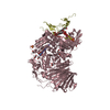
| Structure viewer | Molecule: MolmilJmol/JSmol |
|---|
- Downloads & links
-Download
| PDBx/mmCIF format | 7kb8.cif.gz | 436.4 KB | Display | PDBx/mmCIF format |
|---|---|---|---|---|
| PDB format | pdb7kb8.ent.gz | 332.1 KB | Display | PDB format |
| PDBx/mmJSON format | 7kb8.json.gz | Tree view | PDBx/mmJSON format | |
| Others |  Other downloads Other downloads |
-Validation report
| Arichive directory | https://data.pdbj.org/pub/pdb/validation_reports/kb/7kb8ftp://data.pdbj.org/pub/pdb/validation_reports/kb/7kb8 | HTTPS FTP |
|---|
-Related structure data
| Related structure data | 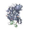 7jtyC  7k9nC 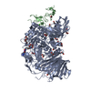 7k9oC 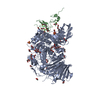 7k9qC  7k9tC 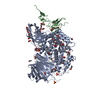 7kadC 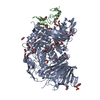 7kb6C 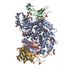 7kbjC  7kbrC 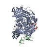 7kryC 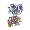 7l9eC 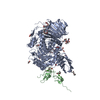 5f0eS S: Starting model for refinement C: citing same article ( |
|---|---|
| Similar structure data |

-Links
 PDBj
PDBj


- Assembly
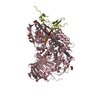
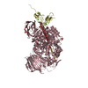
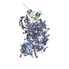
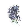
| Deposited unit | 
| |||||||||||||||||||||||||||||||||||||||||||||||||||||||||||||||||||||||||||||||
|---|---|---|---|---|---|---|---|---|---|---|---|---|---|---|---|---|---|---|---|---|---|---|---|---|---|---|---|---|---|---|---|---|---|---|---|---|---|---|---|---|---|---|---|---|---|---|---|---|---|---|---|---|---|---|---|---|---|---|---|---|---|---|---|---|---|---|---|---|---|---|---|---|---|---|---|---|---|---|---|---|
| 1 | 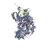
| |||||||||||||||||||||||||||||||||||||||||||||||||||||||||||||||||||||||||||||||
| 2 | 
| |||||||||||||||||||||||||||||||||||||||||||||||||||||||||||||||||||||||||||||||
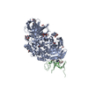
| Unit cell |
| |||||||||||||||||||||||||||||||||||||||||||||||||||||||||||||||||||||||||||||||
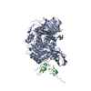
| Noncrystallographic symmetry (NCS) | NCS domain:
NCS domain segments:
|