 Movie
Movie Controller


+ Open data

- Basic information
| Entry | Database: PDB / ID: 7k4i | |||||||||
|---|---|---|---|---|---|---|---|---|---|---|
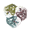
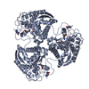
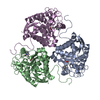
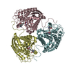
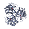
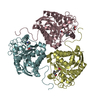
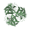
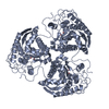
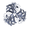
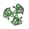
| Title | Human Arginase 1 in complex with compound 06. | |||||||||
 Components Components | Arginase-1 | |||||||||
 Keywords Keywords | HYDROLASE/HYDROLASE inhibitor / Arginase / hydrolase / arginine / urea cycle / inhibitor / HYDROLASE-HYDROLASE inhibitor complex | |||||||||
| Function / homology |  Function and homology information Function and homology informationpositive regulation of neutrophil mediated killing of fungus / Urea cycle / negative regulation of T-helper 2 cell cytokine production / arginase / arginase activity / urea cycle / response to nematode / defense response to protozoan / negative regulation of activated T cell proliferation / negative regulation of type II interferon-mediated signaling pathway ...positive regulation of neutrophil mediated killing of fungus / Urea cycle / negative regulation of T-helper 2 cell cytokine production / arginase / arginase activity / urea cycle / response to nematode / defense response to protozoan / negative regulation of activated T cell proliferation / negative regulation of type II interferon-mediated signaling pathway / L-arginine catabolic process / negative regulation of T cell proliferation / specific granule lumen / azurophil granule lumen / manganese ion binding / adaptive immune response / innate immune response / Neutrophil degranulation / : / extracellular region / nucleus / cytoplasm / cytosol Similarity search - Function | |||||||||
| Biological species |  Homo sapiens (human) Homo sapiens (human) | |||||||||
| Method |  X-RAY DIFFRACTION / SYNCHROTRON / MOLECULAR REPLACEMENT / Resolution: 1.98 Å X-RAY DIFFRACTION / SYNCHROTRON / MOLECULAR REPLACEMENT / Resolution: 1.98 Å | |||||||||
 Authors Authors | Palte, R.L. | |||||||||
 Citation Citation | Journal: Acs Med.Chem.Lett. / Year: 2021 Title: Comprehensive Strategies to Bicyclic Prolines: Applications in the Synthesis of Potent Arginase Inhibitors. Authors: Li, D. / Zhang, H. / Lyons, T.W. / Lu, M. / Achab, A. / Pu, Q. / Childers, M. / Mitcheltree, M.J. / Wang, J. / Martinot, T.A. / McMinn, S.E. / Sloman, D.L. / Palani, A. / Beard, A. / Nogle, ...Authors: Li, D. / Zhang, H. / Lyons, T.W. / Lu, M. / Achab, A. / Pu, Q. / Childers, M. / Mitcheltree, M.J. / Wang, J. / Martinot, T.A. / McMinn, S.E. / Sloman, D.L. / Palani, A. / Beard, A. / Nogle, L. / Gathiaka, S. / Sauri, J. / Kim, H.Y. / Adpressa, D. / Spacciapoli, P. / Miller, J.R. / Palte, R.L. / Lesburg, C.A. / Cumming, J. / Fischer, C. | |||||||||
| History |
|
- Structure visualization
| Structure viewer | Molecule: MolmilJmol/JSmol |
|---|
- Downloads & links
-Download
| PDBx/mmCIF format | 7k4i.cif.gz | 381.3 KB | Display | PDBx/mmCIF format |
|---|---|---|---|---|
| PDB format | pdb7k4i.ent.gz | 308.9 KB | Display | PDB format |
| PDBx/mmJSON format | 7k4i.json.gz | Tree view | PDBx/mmJSON format | |
| Others |  Other downloads Other downloads |
-Validation report
| Arichive directory | https://data.pdbj.org/pub/pdb/validation_reports/k4/7k4iftp://data.pdbj.org/pub/pdb/validation_reports/k4/7k4i | HTTPS FTP |
|---|
-Related structure data
| Related structure data | 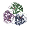 7k4gC 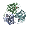 7k4hC  7k4jC 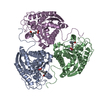 7k4kC 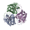 6v7eS S: Starting model for refinement C: citing same article ( |
|---|---|
| Similar structure data |


-Links
 PDBj
PDBj
- Assembly
| Deposited unit | 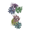
| ||||||||
|---|---|---|---|---|---|---|---|---|---|
| 1 | 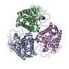
| ||||||||
| 2 | 
| ||||||||
| Unit cell |
|
-Components
| #1: Protein | Mass: 34779.879 Da / Num. of mol.: 6 Source method: isolated from a genetically manipulated source Source: (gene. exp.) Homo sapiens (human) / Gene: ARG1 / Production host:  #2: Chemical | ChemComp-MN /   Mass: 54.938 Da / Num. of mol.: 12 / Source method: obtained synthetically / Formula: Mn Mass: 54.938 Da / Num. of mol.: 12 / Source method: obtained synthetically / Formula: Mn#3: Chemical | ChemComp-VUY /   Mass: 262.111 Da / Num. of mol.: 6 / Source method: obtained synthetically / Formula: C9H17BNO5S / Feature type: SUBJECT OF INVESTIGATION Mass: 262.111 Da / Num. of mol.: 6 / Source method: obtained synthetically / Formula: C9H17BNO5S / Feature type: SUBJECT OF INVESTIGATION#4: Water | ChemComp-HOH / |  Mass: 18.015 Da / Num. of mol.: 812 / Source method: isolated from a natural source / Formula: H2O Mass: 18.015 Da / Num. of mol.: 812 / Source method: isolated from a natural source / Formula: H2OHas ligand of interest | Y | |
|---|
-Experimental details
-Experiment
| Experiment | Method: X-RAY DIFFRACTION / Number of used crystals: 1 |
|---|
- Sample preparation
| Crystal | Density Matthews: 2.48 Å3/Da / Density % sol: 50.41 % |
|---|---|
| Crystal grow | Temperature: 293 K / Method: vapor diffusion, hanging drop / pH: 7 Details: 10% MMT (pH 7.0) 0.1 M ammonium formate, 16-22% PEG 8000 |
-Data collection
| Diffraction | Mean temperature: 100 K / Serial crystal experiment: N |
|---|---|
| Diffraction source | Source: SYNCHROTRON / Site: APS  / Beamline: 17-ID / Wavelength: 1 Å / Beamline: 17-ID / Wavelength: 1 Å |
| Detector | Type: DECTRIS PILATUS 6M / Detector: PIXEL / Date: Nov 29, 2018 |
| Radiation | Protocol: SINGLE WAVELENGTH / Monochromatic (M) / Laue (L): M / Scattering type: x-ray |
| Radiation wavelength | Wavelength: 1 Å / Relative weight: 1 |
| Reflection | Resolution: 1.98→71.78 Å / Num. obs: 125565 / % possible obs: 89.3 % / Redundancy: 3.4 % / Biso Wilson estimate: 26.53 Å2 / CC1/2: 0.994 / Net I/σ(I): 8.6 |
| Reflection shell | Resolution: 1.98→2.09 Å / Num. unique obs: 12199 / CC1/2: 0.758 |
- Processing
| Software |
| ||||||||||||||||||||||||||||||||||||||||||||||||||||||||||||||||||||||||||||||||||||||||||||||||||||||||||||
|---|---|---|---|---|---|---|---|---|---|---|---|---|---|---|---|---|---|---|---|---|---|---|---|---|---|---|---|---|---|---|---|---|---|---|---|---|---|---|---|---|---|---|---|---|---|---|---|---|---|---|---|---|---|---|---|---|---|---|---|---|---|---|---|---|---|---|---|---|---|---|---|---|---|---|---|---|---|---|---|---|---|---|---|---|---|---|---|---|---|---|---|---|---|---|---|---|---|---|---|---|---|---|---|---|---|---|---|---|---|
| Refinement | Method to determine structure: MOLECULAR REPLACEMENT Starting model: 6V7E Resolution: 1.98→71.78 Å / Cor.coef. Fo:Fc: 0.878 / Cor.coef. Fo:Fc free: 0.85 / SU R Cruickshank DPI: 0.227 / Cross valid method: THROUGHOUT / σ(F): 0 / SU R Blow DPI: 0.223 / SU Rfree Blow DPI: 0.179 / SU Rfree Cruickshank DPI: 0.182
| ||||||||||||||||||||||||||||||||||||||||||||||||||||||||||||||||||||||||||||||||||||||||||||||||||||||||||||
| Displacement parameters | Biso max: 130.64 Å2 / Biso mean: 26.75 Å2 / Biso min: 4.97 Å2
| ||||||||||||||||||||||||||||||||||||||||||||||||||||||||||||||||||||||||||||||||||||||||||||||||||||||||||||
| Refine analyze | Luzzati coordinate error obs: 0.32 Å | ||||||||||||||||||||||||||||||||||||||||||||||||||||||||||||||||||||||||||||||||||||||||||||||||||||||||||||
| Refinement step | Cycle: final / Resolution: 1.98→71.78 Å
| ||||||||||||||||||||||||||||||||||||||||||||||||||||||||||||||||||||||||||||||||||||||||||||||||||||||||||||
| Refine LS restraints |
| ||||||||||||||||||||||||||||||||||||||||||||||||||||||||||||||||||||||||||||||||||||||||||||||||||||||||||||
| LS refinement shell | Resolution: 1.98→2.03 Å / Rfactor Rfree error: 0 / Total num. of bins used: 20
|
