 Movie
Movie Controller

[English] 日本語
 Yorodumi
- PDB-3skk: Crystal structure of human arginase I in complex with the inhibit... -
+ Open data

- Basic information
| Entry | Database: PDB / ID: 3skk | ||||||
|---|---|---|---|---|---|---|---|
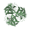
| Title | Crystal structure of human arginase I in complex with the inhibitor FABH, Resolution 1.70 A, twinned structure | ||||||
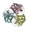
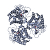
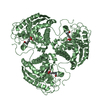
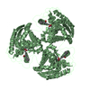
 Components Components | Arginase-1 | ||||||
 Keywords Keywords | HYDROLASE/HYDROLASE INHIBITOR / ABH inhibitor derivative / twinning / HYDROLASE-HYDROLASE INHIBITOR complex | ||||||
| Function / homology |  Function and homology information Function and homology informationpositive regulation of neutrophil mediated killing of fungus / negative regulation of T-helper 2 cell cytokine production / Urea cycle / arginase / arginase activity / urea cycle / response to nematode / defense response to protozoan / negative regulation of activated T cell proliferation / negative regulation of type II interferon-mediated signaling pathway ...positive regulation of neutrophil mediated killing of fungus / negative regulation of T-helper 2 cell cytokine production / Urea cycle / arginase / arginase activity / urea cycle / response to nematode / defense response to protozoan / negative regulation of activated T cell proliferation / negative regulation of type II interferon-mediated signaling pathway / negative regulation of T cell proliferation / L-arginine catabolic process / specific granule lumen / azurophil granule lumen / manganese ion binding / adaptive immune response / innate immune response / Neutrophil degranulation / : / extracellular region / nucleus / cytosol / cytoplasm Similarity search - Function | ||||||
| Biological species |  Homo sapiens (human) Homo sapiens (human) | ||||||
| Method |  X-RAY DIFFRACTION / SYNCHROTRON / MOLECULAR REPLACEMENT / Resolution: 1.701 Å X-RAY DIFFRACTION / SYNCHROTRON / MOLECULAR REPLACEMENT / Resolution: 1.701 Å | ||||||
 Authors Authors | Thorn, K.J. / Di Costanzo, L. / Christianson, D.W. | ||||||
 Citation Citation | Journal: J.Med.Chem. / Year: 2011 Title: Binding of alpha , alpha-disubstituted amino acids to arginase suggests new avenues for inhibitor design. Authors: Ilies, M. / Di Costanzo, L. / Dowling, D.P. / Thorn, K.J. / Christianson, D.W. | ||||||
| History |
|
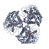
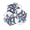
- Structure visualization
| Structure viewer | Molecule: MolmilJmol/JSmol |
|---|
- Downloads & links
-Download
| PDBx/mmCIF format | 3skk.cif.gz | 149.7 KB | Display | PDBx/mmCIF format |
|---|---|---|---|---|
| PDB format | pdb3skk.ent.gz | 115.9 KB | Display | PDB format |
| PDBx/mmJSON format | 3skk.json.gz | Tree view | PDBx/mmJSON format | |
| Others |  Other downloads Other downloads |
-Validation report
| Arichive directory | https://data.pdbj.org/pub/pdb/validation_reports/sk/3skkftp://data.pdbj.org/pub/pdb/validation_reports/sk/3skk | HTTPS FTP |
|---|
-Related structure data
| Related structure data |  3gmzC  3gn0C  3sjtC 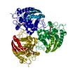 3sl0C  3sl1C 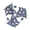 2zavS C: citing same article ( S: Starting model for refinement |
|---|---|
| Similar structure data |


-Links
 PDBj
PDBj
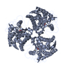
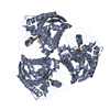
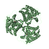
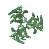
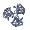
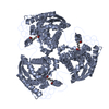
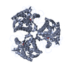
- Assembly
| Deposited unit | 
| |||||||||||||||||||||||||||
|---|---|---|---|---|---|---|---|---|---|---|---|---|---|---|---|---|---|---|---|---|---|---|---|---|---|---|---|---|
| 1 | 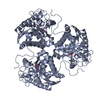
| |||||||||||||||||||||||||||
| 2 | 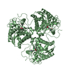
| |||||||||||||||||||||||||||
| Unit cell |
| |||||||||||||||||||||||||||
| Components on special symmetry positions |
|
-Components
| #1: Protein | Mass: 34779.879 Da / Num. of mol.: 2 Source method: isolated from a genetically manipulated source Source: (gene. exp.) Homo sapiens (human) / Gene: ARG1 / Plasmid: pet11d / Production host:  #2: Chemical | ChemComp-MN /   Mass: 54.938 Da / Num. of mol.: 4 / Source method: obtained synthetically / Formula: Mn Mass: 54.938 Da / Num. of mol.: 4 / Source method: obtained synthetically / Formula: Mn#3: Chemical | 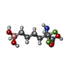  Type: L-peptide linking / Mass: 242.006 Da / Num. of mol.: 2 / Source method: obtained synthetically / Formula: C7H15BF2NO5 Type: L-peptide linking / Mass: 242.006 Da / Num. of mol.: 2 / Source method: obtained synthetically / Formula: C7H15BF2NO5#4: Water | ChemComp-HOH / |  Mass: 18.015 Da / Num. of mol.: 760 / Source method: isolated from a natural source / Formula: H2O Mass: 18.015 Da / Num. of mol.: 760 / Source method: isolated from a natural source / Formula: H2ONonpolymer details | THE STARTING MATERIAL 2-AMINO-6-BORONO-2-(DIFLUOROMETHYL)HEXANOIC ACID (FABH) UNDERGOES TO ...THE STARTING MATERIAL 2-AMINO-6-BORONO-2-(DIFLUOROME | |
|---|
-Experimental details
-Experiment
| Experiment | Method: X-RAY DIFFRACTION / Number of used crystals: 1 |
|---|
- Sample preparation
| Crystal | Density Matthews: 2.34 Å3/Da / Density % sol: 47.36 % |
|---|---|
| Crystal grow | Temperature: 298 K / Method: vapor diffusion, hanging drop / pH: 7.8 Details: 3 uL of protein solution [3.5 mg/mL HAI, 50 mM bicine (pH 8.5), 2 mM FABH, 100 M MnCl2] and 3 uL of precipitant solution [0.1 M HEPES (pH 7.0), 22-28% Jeffamine] were equilibrated against a ...Details: 3 uL of protein solution [3.5 mg/mL HAI, 50 mM bicine (pH 8.5), 2 mM FABH, 100 M MnCl2] and 3 uL of precipitant solution [0.1 M HEPES (pH 7.0), 22-28% Jeffamine] were equilibrated against a 1 mL reservoir of precipitant solution, VAPOR DIFFUSION, HANGING DROP, temperature 298K |
-Data collection
| Diffraction | Mean temperature: 100 K |
|---|---|
| Diffraction source | Source: SYNCHROTRON / Site: APS  / Beamline: 24-ID-E / Wavelength: 1 Å / Beamline: 24-ID-E / Wavelength: 1 Å |
| Detector | Type: ADSC QUANTUM 315 / Detector: CCD |
| Radiation | Protocol: SINGLE WAVELENGTH / Monochromatic (M) / Laue (L): M / Scattering type: x-ray |
| Radiation wavelength | Wavelength: 1 Å / Relative weight: 1 |
| Reflection twin | Operator: -h,-k,l / Fraction: 0.5 |
| Reflection | Resolution: 1.7→50 Å / Num. all: 66860 / Num. obs: 66860 / % possible obs: 99 % / Rmerge(I) obs: 0.148 / Net I/σ(I): 11.6 |
| Reflection shell | Resolution: 1.7→1.8 Å / Rmerge(I) obs: 0.577 / Mean I/σ(I) obs: 2.3 / % possible all: 98.2 |
- Processing
| Software |
| |||||||||||||||||||||||||||||||||||||||||||||||||||||||||||||||||||||||||||||||||||||||||||||||||||||||||||||||||||||||||||||||||||||||||||||||||||
|---|---|---|---|---|---|---|---|---|---|---|---|---|---|---|---|---|---|---|---|---|---|---|---|---|---|---|---|---|---|---|---|---|---|---|---|---|---|---|---|---|---|---|---|---|---|---|---|---|---|---|---|---|---|---|---|---|---|---|---|---|---|---|---|---|---|---|---|---|---|---|---|---|---|---|---|---|---|---|---|---|---|---|---|---|---|---|---|---|---|---|---|---|---|---|---|---|---|---|---|---|---|---|---|---|---|---|---|---|---|---|---|---|---|---|---|---|---|---|---|---|---|---|---|---|---|---|---|---|---|---|---|---|---|---|---|---|---|---|---|---|---|---|---|---|---|---|---|---|
| Refinement | Method to determine structure: MOLECULAR REPLACEMENT Starting model: PDB entry 2ZAV Resolution: 1.701→37.779 Å / σ(F): 0.13 / Phase error: 24.55 / Stereochemistry target values: TWIN_LSQ_F
| |||||||||||||||||||||||||||||||||||||||||||||||||||||||||||||||||||||||||||||||||||||||||||||||||||||||||||||||||||||||||||||||||||||||||||||||||||
| Solvent computation | Shrinkage radii: 0.9 Å / VDW probe radii: 1.11 Å / Solvent model: FLAT BULK SOLVENT MODEL / Bsol: 43.417 Å2 / ksol: 0.324 e/Å3 | |||||||||||||||||||||||||||||||||||||||||||||||||||||||||||||||||||||||||||||||||||||||||||||||||||||||||||||||||||||||||||||||||||||||||||||||||||
| Displacement parameters |
| |||||||||||||||||||||||||||||||||||||||||||||||||||||||||||||||||||||||||||||||||||||||||||||||||||||||||||||||||||||||||||||||||||||||||||||||||||
| Refinement step | Cycle: LAST / Resolution: 1.701→37.779 Å
| |||||||||||||||||||||||||||||||||||||||||||||||||||||||||||||||||||||||||||||||||||||||||||||||||||||||||||||||||||||||||||||||||||||||||||||||||||
| Refine LS restraints |
| |||||||||||||||||||||||||||||||||||||||||||||||||||||||||||||||||||||||||||||||||||||||||||||||||||||||||||||||||||||||||||||||||||||||||||||||||||
| LS refinement shell |
|
