ムービー
ムービー コントローラー


+ データを開く

- 基本情報
基本情報
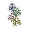
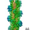
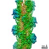
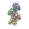
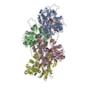
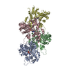
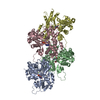
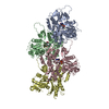
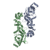
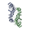
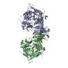
| 登録情報 | データベース: PDB / ID: 7k20 | |||||||||||||||||||||
|---|---|---|---|---|---|---|---|---|---|---|---|---|---|---|---|---|---|---|---|---|---|---|
| タイトル | Cryo-EM structure of pyrene-labeled ADP-actin filaments | |||||||||||||||||||||
 要素 要素 | Actin, alpha skeletal muscle | |||||||||||||||||||||
 キーワード キーワード | CYTOSOLIC PROTEIN / actin / pyrene / fluorescence / ADP | |||||||||||||||||||||
| 機能・相同性 |  機能・相同性情報 機能・相同性情報Striated Muscle Contraction / striated muscle thin filament / skeletal muscle thin filament assembly / skeletal muscle fiber development / stress fiber / actin filament / 加水分解酵素; 酸無水物に作用; 酸無水物に作用・細胞または細胞小器官の運動に関与 / actin cytoskeleton / hydrolase activity / ATP binding 類似検索 - 分子機能 | |||||||||||||||||||||
| 生物種 |  | |||||||||||||||||||||
| 手法 | 電子顕微鏡法 / らせん対称体再構成法 / クライオ電子顕微鏡法 / 解像度: 3.2 Å | |||||||||||||||||||||
 データ登録者 データ登録者 | Chou, S.Z. / Pollard, T.D. | |||||||||||||||||||||
| 資金援助 |  米国, 1件 米国, 1件
| |||||||||||||||||||||
 引用 引用 | ジャーナル: Nat Commun / 年: 2020 タイトル: Cryo-electron microscopy structures of pyrene-labeled ADP-P- and ADP-actin filaments. 著者: Steven Z Chou / Thomas D Pollard / 要旨: Since the fluorescent reagent N-(1-pyrene)iodoacetamide was first used to label skeletal muscle actin in 1981, the pyrene-labeled actin has become the most widely employed tool to measure the ...Since the fluorescent reagent N-(1-pyrene)iodoacetamide was first used to label skeletal muscle actin in 1981, the pyrene-labeled actin has become the most widely employed tool to measure the kinetics of actin polymerization and the interaction between actin and actin-binding proteins. Here we report high-resolution cryo-electron microscopy structures of actin filaments with N-1-pyrene conjugated to cysteine 374 and either ADP (3.2 Å) or ADP-phosphate (3.0 Å) in the active site. Polymerization buries pyrene in a hydrophobic cavity between subunits along the long-pitch helix with only minor differences in conformation compared with native actin filaments. These structures explain how polymerization increases the fluorescence 20-fold, how myosin and cofilin binding to filaments reduces the fluorescence, and how profilin binding to actin monomers increases the fluorescence. | |||||||||||||||||||||
| 履歴 |
|
- 構造の表示
構造の表示
| ムービー |
ムービービューア |
|---|---|
| 構造ビューア | 分子: MolmilJmol/JSmol |
- ダウンロードとリンク
-ダウンロード
| PDBx/mmCIF形式 | 7k20.cif.gz | 263 KB | 表示 | PDBx/mmCIF形式 |
|---|---|---|---|---|
| PDB形式 | pdb7k20.ent.gz | 214.8 KB | 表示 | PDB形式 |
| PDBx/mmJSON形式 | 7k20.json.gz | ツリー表示 | PDBx/mmJSON形式 | |
| その他 |  その他のダウンロード その他のダウンロード |
-検証レポート
| 文書・要旨 | 7k20_validation.pdf.gz | 1.1 MB | 表示 | wwPDB検証レポート |
|---|---|---|---|---|
| 文書・詳細版 | 7k20_full_validation.pdf.gz | 1.1 MB | 表示 | |
| XML形式データ | 7k20_validation.xml.gz | 44.5 KB | 表示 | |
| CIF形式データ | 7k20_validation.cif.gz | 66.4 KB | 表示 | |
| アーカイブディレクトリ | https://data.pdbj.org/pub/pdb/validation_reports/k2/7k20ftp://data.pdbj.org/pub/pdb/validation_reports/k2/7k20 | HTTPS FTP |
-関連構造データ


-リンク
 PDBj
PDBj


- 集合体
集合体
| 登録構造単位 | 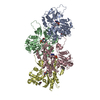
|
|---|---|
| 1 |
|
-要素
| #1: タンパク質 | 分子量: 41875.633 Da / 分子数: 4 / 由来タイプ: 天然 詳細: The C19 atom of pyrene (1T4) is chemically conjugated to the SG atom of actin C374 由来: (天然) #2: 化合物 | ChemComp-MG /   分子量: 24.305 Da / 分子数: 4 / 由来タイプ: 合成 / 式: Mg 分子量: 24.305 Da / 分子数: 4 / 由来タイプ: 合成 / 式: Mg#3: 化合物 | ChemComp-ADP /   分子量: 427.201 Da / 分子数: 4 / 由来タイプ: 合成 / 式: C10H15N5O10P2 / タイプ: SUBJECT OF INVESTIGATION / コメント: ADP, エネルギー貯蔵分子*YM 分子量: 427.201 Da / 分子数: 4 / 由来タイプ: 合成 / 式: C10H15N5O10P2 / タイプ: SUBJECT OF INVESTIGATION / コメント: ADP, エネルギー貯蔵分子*YM#4: 化合物 | ChemComp-1T4 /   分子量: 259.302 Da / 分子数: 4 / 由来タイプ: 合成 / 式: C18H13NO / タイプ: SUBJECT OF INVESTIGATION 分子量: 259.302 Da / 分子数: 4 / 由来タイプ: 合成 / 式: C18H13NO / タイプ: SUBJECT OF INVESTIGATION研究の焦点であるリガンドがあるか | Y | Has protein modification | Y | |
|---|
-実験情報
-実験
| 実験 | 手法: 電子顕微鏡法 |
|---|---|
| EM実験 | 試料の集合状態: FILAMENT / 3次元再構成法: らせん対称体再構成法 |
- 試料調製
試料調製
| 構成要素 | 名称: Pyrene-labeled ADP-actin filaments / タイプ: COMPLEX 詳細: Pyene is chemically conjugated to the side chain of actin C374 Entity ID: #1 / 由来: NATURAL |
|---|---|
| 由来(天然) | 生物種: |
| 緩衝液 | pH: 7 |
| 試料 | 包埋: NO / シャドウイング: NO / 染色: NO / 凍結: YES |
| 試料支持 | 詳細: unspecified |
| 急速凍結 | 装置: FEI VITROBOT MARK IV / 凍結剤: ETHANE / 湿度: 100 % / 凍結前の試料温度: 283 K |
- 電子顕微鏡撮影
| 実験機器 |  モデル: Titan Krios / 画像提供: FEI Company |
|---|---|
| 顕微鏡 | モデル: FEI TITAN KRIOS |
| 電子銃 | 電子線源:  FIELD EMISSION GUN / 加速電圧: 300 kV / 照射モード: FLOOD BEAM FIELD EMISSION GUN / 加速電圧: 300 kV / 照射モード: FLOOD BEAM |
| 電子レンズ | モード: BRIGHT FIELD |
| 撮影 | 平均露光時間: 11 sec. / 電子線照射量: 67.9 e/Å2 / 検出モード: SUPER-RESOLUTION フィルム・検出器のモデル: GATAN K2 QUANTUM (4k x 4k) 撮影したグリッド数: 1 |
- 解析
解析
| ソフトウェア | 名称: PHENIX / バージョン: 1.12_2829: / 分類: 精密化 | ||||||||||||||||||||||||
|---|---|---|---|---|---|---|---|---|---|---|---|---|---|---|---|---|---|---|---|---|---|---|---|---|---|
| EMソフトウェア |
| ||||||||||||||||||||||||
| CTF補正 | タイプ: PHASE FLIPPING AND AMPLITUDE CORRECTION | ||||||||||||||||||||||||
| らせん対称 | 回転角度/サブユニット: -166.57869 ° / 軸方向距離/サブユニット: 27.385195 Å / らせん対称軸の対称性: C1 | ||||||||||||||||||||||||
| 3次元再構成 | 解像度: 3.2 Å / 解像度の算出法: FSC 0.143 CUT-OFF / 粒子像の数: 268618 / 対称性のタイプ: HELICAL | ||||||||||||||||||||||||
| 原子モデル構築 | プロトコル: OTHER / 空間: REAL 詳細: Restraints for pyrene were generated with eLBOW in Phenix. The coordinates of actin and pyrene were joined together manually in a text editor, and then fitted into the map in Coot. | ||||||||||||||||||||||||
| 原子モデル構築 | PDB-ID: 6DJN Accession code: 6DJN / Source name: PDB / タイプ: experimental model | ||||||||||||||||||||||||
| 拘束条件 |
|