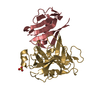
 Movie
Movie Controller


+ Open data

- Basic information
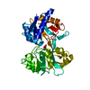
| Entry | Database: PDB / ID: 7jys | ||||||
|---|---|---|---|---|---|---|---|
| Title | hALK in complex with 3-(3-chlorophenyl)-5-methyl-1H-pyrazole | ||||||
 Components Components | ALK tyrosine kinase receptor | ||||||
 Keywords Keywords | TRANSFERASE / ALK tyrosine kinase receptor / INHIBITOR / COMPLEX / KINASE / SBDD / TRANSFERASE-INHIBITOR COMPLEX | ||||||
| Function / homology |  Function and homology information Function and homology informationASP-3026-resistant ALK mutants / NVP-TAE684-resistant ALK mutants / alectinib-resistant ALK mutants / brigatinib-resistant ALK mutants / ceritinib-resistant ALK mutants / crizotinib-resistant ALK mutants / lorlatinib-resistant ALK mutants / MDK and PTN in ALK signaling / receptor signaling protein tyrosine kinase activator activity / regulation of dopamine receptor signaling pathway ...ASP-3026-resistant ALK mutants / NVP-TAE684-resistant ALK mutants / alectinib-resistant ALK mutants / brigatinib-resistant ALK mutants / ceritinib-resistant ALK mutants / crizotinib-resistant ALK mutants / lorlatinib-resistant ALK mutants / MDK and PTN in ALK signaling / receptor signaling protein tyrosine kinase activator activity / regulation of dopamine receptor signaling pathway / response to environmental enrichment / ALK mutants bind TKIs / peptidyl-tyrosine autophosphorylation / phosphorylation / positive regulation of dendrite development / regulation of neuron differentiation / Signaling by ALK / adult behavior / response to stress / negative regulation of lipid catabolic process / neuron development / energy homeostasis / swimming behavior / transmembrane receptor protein tyrosine kinase activity / cell surface receptor protein tyrosine kinase signaling pathway / : / hippocampus development / receptor protein-tyrosine kinase / Signaling by ALK fusions and activated point mutants / protein autophosphorylation / regulation of cell population proliferation / heparin binding / protein tyrosine kinase activity / regulation of apoptotic process / signaling receptor complex / signal transduction / protein-containing complex / extracellular exosome / ATP binding / identical protein binding / plasma membrane Similarity search - Function | ||||||
| Biological species |  Homo sapiens (human) Homo sapiens (human) | ||||||
| Method |  X-RAY DIFFRACTION / SYNCHROTRON / MOLECULAR REPLACEMENT / Resolution: 2.22 Å X-RAY DIFFRACTION / SYNCHROTRON / MOLECULAR REPLACEMENT / Resolution: 2.22 Å | ||||||
 Authors Authors | McGrath, A.P. / Zou, H. / Lane, W. / Saikatendu, K. | ||||||
 Citation Citation | Journal: Acs Omega / Year: 2020 Title: Discovery of Novel and Highly Selective Cyclopropane ALK Inhibitors through a Fragment-Assisted, Structure-Based Drug Design. Authors: Fujimori, I. / Wakabayashi, T. / Murakami, M. / Okabe, A. / Ishii, T. / McGrath, A. / Zou, H. / Saikatendu, K.S. / Imoto, H. | ||||||
| History |
|
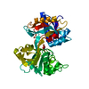
- Structure visualization
| Structure viewer | Molecule: MolmilJmol/JSmol |
|---|
- Downloads & links
-Download
| PDBx/mmCIF format | 7jys.cif.gz | 146.6 KB | Display | PDBx/mmCIF format |
|---|---|---|---|---|
| PDB format | pdb7jys.ent.gz | 93.3 KB | Display | PDB format |
| PDBx/mmJSON format | 7jys.json.gz | Tree view | PDBx/mmJSON format | |
| Others |  Other downloads Other downloads |
-Validation report
| Arichive directory | https://data.pdbj.org/pub/pdb/validation_reports/jy/7jysftp://data.pdbj.org/pub/pdb/validation_reports/jy/7jys | HTTPS FTP |
|---|
-Related structure data
| Related structure data |  7jy4C  7jyrC  7jytC 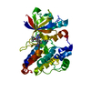 4z55S C: citing same article ( S: Starting model for refinement |
|---|---|
| Similar structure data |


-Links
 PDBj
PDBj


- Assembly
| Deposited unit | 
| ||||||||||||
|---|---|---|---|---|---|---|---|---|---|---|---|---|---|
| 1 |
| ||||||||||||
| Unit cell |
|
-Components
| #1: Protein | Mass: 36045.371 Da / Num. of mol.: 1 Source method: isolated from a genetically manipulated source Source: (gene. exp.) Homo sapiens (human) / Gene: ALK / Plasmid: pFastBacHTb / Cell (production host): Sf9 / Production host:   Spodoptera frugiperda (fall armyworm) Spodoptera frugiperda (fall armyworm)References: UniProt: Q9UM73, receptor protein-tyrosine kinase |
|---|---|
| #2: Chemical | ChemComp-VTD / 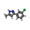  Mass: 192.645 Da / Num. of mol.: 1 / Source method: obtained synthetically / Formula: C10H9ClN2 / Feature type: SUBJECT OF INVESTIGATION Mass: 192.645 Da / Num. of mol.: 1 / Source method: obtained synthetically / Formula: C10H9ClN2 / Feature type: SUBJECT OF INVESTIGATION |
| #3: Chemical | ChemComp-EDO /   Mass: 62.068 Da / Num. of mol.: 1 / Source method: obtained synthetically / Formula: C2H6O2 Mass: 62.068 Da / Num. of mol.: 1 / Source method: obtained synthetically / Formula: C2H6O2 |
| #4: Water | ChemComp-HOH /  Mass: 18.015 Da / Num. of mol.: 55 / Source method: isolated from a natural source / Formula: H2O Mass: 18.015 Da / Num. of mol.: 55 / Source method: isolated from a natural source / Formula: H2O |
| Has ligand of interest | Y |
-Experimental details
-Experiment
| Experiment | Method: X-RAY DIFFRACTION / Number of used crystals: 1 |
|---|
- Sample preparation
| Crystal | Density Matthews: 2.11 Å3/Da / Density % sol: 41.81 % |
|---|---|
| Crystal grow | Temperature: 293 K / Method: vapor diffusion / pH: 8 Details: 25 mM Tris pH 8.0, 150 mM NaCl, 0.5 mM EDTA, 0.25 mM TCEP |
-Data collection
| Diffraction | Mean temperature: 100 K / Serial crystal experiment: N |
|---|---|
| Diffraction source | Source: SYNCHROTRON / Site: ALS  / Beamline: 5.0.3 / Wavelength: 0.976 Å / Beamline: 5.0.3 / Wavelength: 0.976 Å |
| Detector | Type: ADSC QUANTUM 315r / Detector: CCD / Date: Oct 5, 2012 |
| Radiation | Protocol: SINGLE WAVELENGTH / Monochromatic (M) / Laue (L): M / Scattering type: x-ray |
| Radiation wavelength | Wavelength: 0.976 Å / Relative weight: 1 |
| Reflection | Resolution: 2.22→29.51 Å / Num. obs: 15566 / % possible obs: 99.13 % / Redundancy: 6.1 % / Biso Wilson estimate: 43.31 Å2 / Rmerge(I) obs: 0.098 / Net I/σ(I): 16.16 |
| Reflection shell | Resolution: 2.22→2.299 Å / Rmerge(I) obs: 0.897 / Mean I/σ(I) obs: 2.36 / Num. unique obs: 1458 / % possible all: 94.97 |
- Processing
| Software |
| |||||||||||||||||||||||||||||||||||||||||||||||||
|---|---|---|---|---|---|---|---|---|---|---|---|---|---|---|---|---|---|---|---|---|---|---|---|---|---|---|---|---|---|---|---|---|---|---|---|---|---|---|---|---|---|---|---|---|---|---|---|---|---|---|
| Refinement | Method to determine structure: MOLECULAR REPLACEMENT Starting model: 4z55 Resolution: 2.22→29.51 Å / SU ML: 0.2686 / Cross valid method: FREE R-VALUE / σ(F): 1.34 / Phase error: 25.71 Stereochemistry target values: GeoStd + Monomer Library + CDL v1.2
| |||||||||||||||||||||||||||||||||||||||||||||||||
| Solvent computation | Shrinkage radii: 0.9 Å / VDW probe radii: 1.11 Å / Solvent model: FLAT BULK SOLVENT MODEL | |||||||||||||||||||||||||||||||||||||||||||||||||
| Displacement parameters | Biso mean: 55.83 Å2 | |||||||||||||||||||||||||||||||||||||||||||||||||
| Refinement step | Cycle: LAST / Resolution: 2.22→29.51 Å
| |||||||||||||||||||||||||||||||||||||||||||||||||
| Refine LS restraints |
| |||||||||||||||||||||||||||||||||||||||||||||||||
| LS refinement shell |
|
