 Movie
Movie Controller

[English] 日本語
 Yorodumi
- PDB-7jyp: Structure of thioredoxin reductase from the thermophilic eubacter... -
+ Open data

- Basic information
| Entry | Database: PDB / ID: 7jyp | ||||||
|---|---|---|---|---|---|---|---|
| Title | Structure of thioredoxin reductase from the thermophilic eubacterium Thermosipho africanus TCF52B | ||||||
 Components Components | Thioredoxin reductase | ||||||
 Keywords Keywords | OXIDOREDUCTASE / Thioredoxin reductase / thermophile / Thermosipho africanus / protein stability / catalysis / NADH | ||||||
| Function / homology |  Function and homology information Function and homology informationthioredoxin-disulfide reductase (NADPH) / thioredoxin-disulfide reductase (NADPH) activity / removal of superoxide radicals / nucleotide binding / cytoplasm Similarity search - Function | ||||||
| Biological species |  Thermosipho africanus (bacteria) Thermosipho africanus (bacteria) | ||||||
| Method |  X-RAY DIFFRACTION / SYNCHROTRON / MOLECULAR REPLACEMENT / Resolution: 1.6 Å X-RAY DIFFRACTION / SYNCHROTRON / MOLECULAR REPLACEMENT / Resolution: 1.6 Å | ||||||
 Authors Authors | Sahtout, N. / Kuttiyatveetil, J.R.A. / Sanders, D.A.R. | ||||||
| Funding support |  Canada, 1items Canada, 1items
| ||||||
 Citation Citation | Journal: To be Published Title: Structure of thioredoxin reductase from the thermophilic eubacterium Thermosipho africanus TCF52B. Authors: Sahtout, N. / Kuttiyatveetil, J.R.A. / Sanders, D.A.R. #1: Journal: Biochim Biophys Acta Proteins Proteom / Year: 2019Title: Structure and function of the putative thioredoxin 1 from the thermophilic eubacterium Thermosipho africanus strain TCF52B. Authors: Sahtout, N. / Kuttiyatveetil, J.R.A. / Sanders, D.A.R. #2: Journal: Acta Crystallogr F Struct Biol Commun / Year: 2016Title: Putative thioredoxin Trx1 from Thermosipho africanus strain TCF52B: expression, purification and structural determination using S-SAD. Authors: Sahtout, N. / Kuttiyatveetil, J.R. / Fodje, M. / Sanders, D.A. | ||||||
| History |
|
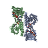
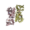
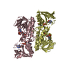
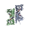
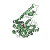
- Structure visualization
| Structure viewer | Molecule: MolmilJmol/JSmol |
|---|
- Downloads & links
-Download
| PDBx/mmCIF format | 7jyp.cif.gz | 79.2 KB | Display | PDBx/mmCIF format |
|---|---|---|---|---|
| PDB format | pdb7jyp.ent.gz | 54.6 KB | Display | PDB format |
| PDBx/mmJSON format | 7jyp.json.gz | Tree view | PDBx/mmJSON format | |
| Others |  Other downloads Other downloads |
-Validation report
| Arichive directory | https://data.pdbj.org/pub/pdb/validation_reports/jy/7jypftp://data.pdbj.org/pub/pdb/validation_reports/jy/7jyp | HTTPS FTP |
|---|
-Related structure data
| Similar structure data |
|---|


-Links
 PDBj
PDBj


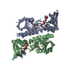
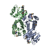
- Assembly
| Deposited unit | 
| ||||||||
|---|---|---|---|---|---|---|---|---|---|
| 1 | 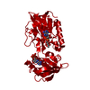
| ||||||||
| Unit cell |
|
-Components
| #1: Protein | Mass: 37443.570 Da / Num. of mol.: 1 Source method: isolated from a genetically manipulated source Source: (gene. exp.) Thermosipho africanus (strain TCF52B) (bacteria)Strain: TCF52B / Gene: trxB, THA_1385 / Plasmid: pEHISTEV / Production host: References: UniProt: B7ICV3, thioredoxin-disulfide reductase (NADPH) |
|---|---|
| #2: Chemical | ChemComp-FAD /   Mass: 785.550 Da / Num. of mol.: 1 / Source method: obtained synthetically / Formula: C27H33N9O15P2 / Comment: FAD*YM Mass: 785.550 Da / Num. of mol.: 1 / Source method: obtained synthetically / Formula: C27H33N9O15P2 / Comment: FAD*YM |
| #3: Chemical | ChemComp-NAD /   Mass: 663.425 Da / Num. of mol.: 1 / Source method: obtained synthetically / Formula: C21H27N7O14P2 / Comment: NAD*YM Mass: 663.425 Da / Num. of mol.: 1 / Source method: obtained synthetically / Formula: C21H27N7O14P2 / Comment: NAD*YM |
| #4: Water | ChemComp-HOH /  Mass: 18.015 Da / Num. of mol.: 93 / Source method: isolated from a natural source / Formula: H2O Mass: 18.015 Da / Num. of mol.: 93 / Source method: isolated from a natural source / Formula: H2O |
| Has ligand of interest | N |
| Has protein modification | Y |
-Experimental details
-Experiment
| Experiment | Method: X-RAY DIFFRACTION / Number of used crystals: 1 |
|---|
- Sample preparation
| Crystal | Density Matthews: 2.13 Å3/Da / Density % sol: 42.15 % / Description: yellow rhomboid shaped crystals |
|---|---|
| Crystal grow | Temperature: 289 K / Method: microbatch Details: 0.2 M Ammonium Chloride, 20 % (w/v) PEG 3350, 0.1 M b-Nicotinamide adenine dinucleotide (NAD) hydrate |
-Data collection
| Diffraction | Mean temperature: 100 K / Serial crystal experiment: N |
|---|---|
| Diffraction source | Source: SYNCHROTRON / Site: CLSI / Beamline: 08ID-1 / Wavelength: 0.9795 Å |
| Detector | Type: DECTRIS PILATUS3 S 6M / Detector: PIXEL / Date: Apr 20, 2016 |
| Radiation | Protocol: SINGLE WAVELENGTH / Monochromatic (M) / Laue (L): M / Scattering type: x-ray |
| Radiation wavelength | Wavelength: 0.9795 Å / Relative weight: 1 |
| Reflection | Resolution: 1.6→48.12 Å / Num. obs: 42842 / % possible obs: 99.2 % / Redundancy: 9.2 % / Biso Wilson estimate: 25.76 Å2 / CC1/2: 0.999 / Net I/σ(I): 11 |
| Reflection shell | Resolution: 1.6→1.657 Å / Num. unique obs: 3980 / CC1/2: 0.17 / % possible all: 93.5 |
- Processing
| Software |
| ||||||||||||||||||||||||||||||||||||||||||||||||||||||||||||||||||||||||||||||||||||||||||||||||||||||||||||||||
|---|---|---|---|---|---|---|---|---|---|---|---|---|---|---|---|---|---|---|---|---|---|---|---|---|---|---|---|---|---|---|---|---|---|---|---|---|---|---|---|---|---|---|---|---|---|---|---|---|---|---|---|---|---|---|---|---|---|---|---|---|---|---|---|---|---|---|---|---|---|---|---|---|---|---|---|---|---|---|---|---|---|---|---|---|---|---|---|---|---|---|---|---|---|---|---|---|---|---|---|---|---|---|---|---|---|---|---|---|---|---|---|---|---|
| Refinement | Method to determine structure: MOLECULAR REPLACEMENT Starting model: Automated Resolution: 1.6→48.119 Å / SU ML: 0.36 / Cross valid method: THROUGHOUT / σ(F): 1.33 / Phase error: 35.67 / Stereochemistry target values: ML
| ||||||||||||||||||||||||||||||||||||||||||||||||||||||||||||||||||||||||||||||||||||||||||||||||||||||||||||||||
| Solvent computation | Shrinkage radii: 0.9 Å / VDW probe radii: 1.11 Å / Solvent model: FLAT BULK SOLVENT MODEL | ||||||||||||||||||||||||||||||||||||||||||||||||||||||||||||||||||||||||||||||||||||||||||||||||||||||||||||||||
| Refinement step | Cycle: LAST / Resolution: 1.6→48.119 Å
| ||||||||||||||||||||||||||||||||||||||||||||||||||||||||||||||||||||||||||||||||||||||||||||||||||||||||||||||||
| Refine LS restraints |
| ||||||||||||||||||||||||||||||||||||||||||||||||||||||||||||||||||||||||||||||||||||||||||||||||||||||||||||||||
| LS refinement shell |
|
