 Movie
Movie Controller

[English] 日本語
 Yorodumi
- PDB-3f8d: Structure of Sulfolobus solfataricus Thioredoxin reductase Mutant... -
+ Open data

- Basic information
| Entry | Database: PDB / ID: 3f8d | ||||||
|---|---|---|---|---|---|---|---|
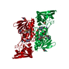
| Title | Structure of Sulfolobus solfataricus Thioredoxin reductase Mutant C147A | ||||||
 Components Components | Thioredoxin reductase (TrxB-3) | ||||||
 Keywords Keywords | OXIDOREDUCTASE / Redox protein / nucleotide binding / FAD / Flavoprotein | ||||||
| Function / homology |  Function and homology information Function and homology informationNADH dehydrogenase / thioredoxin-disulfide reductase (NADPH) / thioredoxin-disulfide reductase (NADPH) activity / removal of superoxide radicals / cell redox homeostasis / nucleotide binding / cytoplasm Similarity search - Function | ||||||
| Biological species |   Sulfolobus solfataricus (archaea) Sulfolobus solfataricus (archaea) | ||||||
| Method |  X-RAY DIFFRACTION / SYNCHROTRON / MOLECULAR REPLACEMENT / Resolution: 1.4 Å X-RAY DIFFRACTION / SYNCHROTRON / MOLECULAR REPLACEMENT / Resolution: 1.4 Å | ||||||
 Authors Authors | Ruggiero, A. / Masullo, M. / Ruocco, M.R. / Arcari, P. / Zagari, A. / Vitagliano, L. | ||||||
 Citation Citation | Journal: Biochim.Biophys.Acta / Year: 2009 Title: Structure and stability of a thioredoxin reductase from Sulfolobus solfataricus: a thermostable protein with two functions Authors: Ruggiero, A. / Masullo, M. / Ruocco, M.R. / Grimaldi, P. / Lanzotti, M.A. / Arcari, P. / Zagari, A. / Vitagliano, L. | ||||||
| History |
|
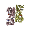
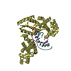
- Structure visualization
| Structure viewer | Molecule: MolmilJmol/JSmol |
|---|
- Downloads & links
-Download
| PDBx/mmCIF format | 3f8d.cif.gz | 253.6 KB | Display | PDBx/mmCIF format |
|---|---|---|---|---|
| PDB format | pdb3f8d.ent.gz | 203.7 KB | Display | PDB format |
| PDBx/mmJSON format | 3f8d.json.gz | Tree view | PDBx/mmJSON format | |
| Others |  Other downloads Other downloads |
-Validation report
| Arichive directory | https://data.pdbj.org/pub/pdb/validation_reports/f8/3f8dftp://data.pdbj.org/pub/pdb/validation_reports/f8/3f8d | HTTPS FTP |
|---|
-Related structure data
| Related structure data |  3f8pSC  3f8rC S: Starting model for refinement C: citing same article ( |
|---|---|
| Similar structure data |


-Links
 PDBj
PDBj


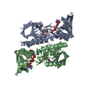
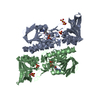
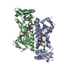
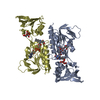
- Assembly
| Deposited unit | 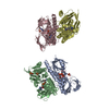
| ||||||||
|---|---|---|---|---|---|---|---|---|---|
| 1 | 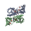
| ||||||||
| 2 | 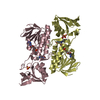
| ||||||||
| Unit cell |
|
-Components
| #1: Protein | Mass: 35234.613 Da / Num. of mol.: 4 / Mutation: C147A Source method: isolated from a genetically manipulated source Source: (gene. exp.) Sulfolobus solfataricus (archaea) / Plasmid: pET-28 / Production host:  References: UniProt: Q97W27, UniProt: Q8X236*PLUS, EC: 1.6.4.5 #2: Chemical | ChemComp-FAD /   Mass: 785.550 Da / Num. of mol.: 4 / Source method: obtained synthetically / Formula: C27H33N9O15P2 / Comment: FAD*YM Mass: 785.550 Da / Num. of mol.: 4 / Source method: obtained synthetically / Formula: C27H33N9O15P2 / Comment: FAD*YM#3: Chemical | ChemComp-SO4 /   Mass: 96.063 Da / Num. of mol.: 6 / Source method: obtained synthetically / Formula: SO4 Mass: 96.063 Da / Num. of mol.: 6 / Source method: obtained synthetically / Formula: SO4#4: Chemical | ChemComp-ACY / |   Mass: 60.052 Da / Num. of mol.: 1 / Source method: obtained synthetically / Formula: C2H4O2 Mass: 60.052 Da / Num. of mol.: 1 / Source method: obtained synthetically / Formula: C2H4O2#5: Water | ChemComp-HOH / |  Mass: 18.015 Da / Num. of mol.: 393 / Source method: isolated from a natural source / Formula: H2O Mass: 18.015 Da / Num. of mol.: 393 / Source method: isolated from a natural source / Formula: H2O |
|---|
-Experimental details
-Experiment
| Experiment | Method: X-RAY DIFFRACTION / Number of used crystals: 1 |
|---|
- Sample preparation
| Crystal | Density Matthews: 2.1 Å3/Da / Density % sol: 41.43 % |
|---|---|
| Crystal grow | Temperature: 293 K / Method: vapor diffusion / pH: 7.5 Details: 12-15% (w/v) poly-ethylene glycol 2000 monomethyl ether, 0.1M (NH4)2SO4, 50mM sodium acetate (pH 4.6), protein 4-9 mg/ml, VAPOR DIFFUSION, temperature 293K |
-Data collection
| Diffraction | Mean temperature: 100 K |
|---|---|
| Diffraction source | Source: SYNCHROTRON / Site: ESRF  / Beamline: ID29 / Wavelength: 0.9756 Å / Beamline: ID29 / Wavelength: 0.9756 Å |
| Detector | Type: ADSC QUANTUM 4 / Detector: CCD / Date: Dec 8, 2003 |
| Radiation | Monochromator: Monochromator / Protocol: SINGLE WAVELENGTH / Monochromatic (M) / Laue (L): M / Scattering type: x-ray |
| Radiation wavelength | Wavelength: 0.9756 Å / Relative weight: 1 |
| Reflection | Resolution: 1.4→30 Å / Num. all: 231790 / Num. obs: 231790 / % possible obs: 98.9 % / Observed criterion σ(F): 0 / Observed criterion σ(I): 0 / Rmerge(I) obs: 0.081 |
| Reflection shell | Resolution: 1.4→1.45 Å / Rmerge(I) obs: 0.326 / % possible all: 91.7 |
- Processing
| Software |
| ||||||||||||||||||||
|---|---|---|---|---|---|---|---|---|---|---|---|---|---|---|---|---|---|---|---|---|---|
| Refinement | Method to determine structure: MOLECULAR REPLACEMENT Starting model: PDB ENTRY 3F8P Resolution: 1.4→15 Å / Cross valid method: THROUGHOUT / σ(F): 0 / Stereochemistry target values: Engh & Huber
| ||||||||||||||||||||
| Refinement step | Cycle: LAST / Resolution: 1.4→15 Å
|
