 Movie
Movie Controller

[English] 日本語
 Yorodumi
- PDB-7jtx: the structural basis of PTEN regulation by multi-site phosphorylation -
+ Open data

- Basic information
| Entry | Database: PDB / ID: 7jtx | ||||||||||||
|---|---|---|---|---|---|---|---|---|---|---|---|---|---|
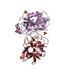
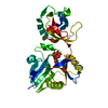
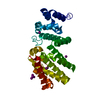
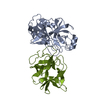
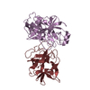
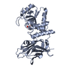
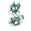
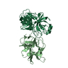
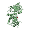
| Title | the structural basis of PTEN regulation by multi-site phosphorylation | ||||||||||||
 Components Components | Phosphatidylinositol 3,4,5-trisphosphate 3-phosphatase and dual-specificity protein phosphatase PTEN | ||||||||||||
 Keywords Keywords | HYDROLASE / PTEN / phosphatase / cellular localization | ||||||||||||
| Biological species |  Homo sapiens (human) Homo sapiens (human) | ||||||||||||
| Method |  X-RAY DIFFRACTION / SYNCHROTRON / MOLECULAR REPLACEMENT / Resolution: 3.23 Å X-RAY DIFFRACTION / SYNCHROTRON / MOLECULAR REPLACEMENT / Resolution: 3.23 Å | ||||||||||||
 Authors Authors | Park, E. / Dempsey, D.R. / Cole, P. | ||||||||||||
| Funding support |  United States, 3items United States, 3items
| ||||||||||||
 Citation Citation | Journal: Nat.Struct.Mol.Biol. / Year: 2021 Title: The structural basis of PTEN regulation by multi-site phosphorylation. Authors: Dempsey, D.R. / Viennet, T. / Iwase, R. / Park, E. / Henriquez, S. / Chen, Z. / Jeliazkov, J.R. / Palanski, B.A. / Phan, K.L. / Coote, P. / Gray, J.J. / Eck, M.J. / Gabelli, S.B. / Arthanari, H. / Cole, P.A. | ||||||||||||
| History |
|
- Structure visualization
| Structure viewer | Molecule:  MolmilJmol/JSmol MolmilJmol/JSmol |
|---|
- Downloads & links
-Download
| PDBx/mmCIF format | 7jtx.cif.gz | 73.9 KB | Display | PDBx/mmCIF format |
|---|---|---|---|---|
| PDB format | pdb7jtx.ent.gz | 51.7 KB | Display | PDB format |
| PDBx/mmJSON format | 7jtx.json.gz | Tree view | PDBx/mmJSON format | |
| Others |  Other downloads Other downloads |
-Validation report
| Arichive directory | https://data.pdbj.org/pub/pdb/validation_reports/jt/7jtxftp://data.pdbj.org/pub/pdb/validation_reports/jt/7jtx | HTTPS FTP |
|---|
-Related structure data
| Related structure data | 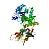 7jukC 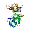 7julC 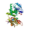 7jvxC  1d5rS S: Starting model for refinement C: citing same article ( |
|---|---|
| Similar structure data |

-Links
 PDBj
PDBj- Assembly
| Deposited unit | 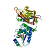
| ||||||||||
|---|---|---|---|---|---|---|---|---|---|---|---|
| 1 |
| ||||||||||
| Unit cell |
|
-Components
| #1: Protein | Mass: 42338.828 Da / Num. of mol.: 1 Source method: isolated from a genetically manipulated source Source: (gene. exp.) Homo sapiens (human) / Production host:   Spodoptera frugiperda (fall armyworm) Spodoptera frugiperda (fall armyworm)References: protein-serine/threonine phosphatase, protein-tyrosine-phosphatase, phosphatidylinositol-3,4,5-trisphosphate 3-phosphatase |
|---|
-Experimental details
-Experiment
| Experiment | Method: X-RAY DIFFRACTION / Number of used crystals: 1 |
|---|
- Sample preparation
| Crystal | Density Matthews: 2.25 Å3/Da / Density % sol: 45.33 % |
|---|---|
| Crystal grow | Temperature: 298 K / Method: vapor diffusion, hanging drop / Details: 300 mM sodium cistrate tribasic, 20% PEG 3350 / Temp details: room temperature |
-Data collection
| Diffraction | Mean temperature: 100 K / Serial crystal experiment: N |
|---|---|
| Diffraction source | Source: SYNCHROTRON / Site: APS / Beamline: 24-ID-E / Wavelength: 0.9798 Å |
| Detector | Type: DECTRIS EIGER X 16M / Detector: PIXEL / Date: Jun 13, 2018 |
| Radiation | Protocol: SINGLE WAVELENGTH / Monochromatic (M) / Laue (L): M / Scattering type: x-ray |
| Radiation wavelength | Wavelength: 0.9798 Å / Relative weight: 1 |
| Reflection | Resolution: 3.05→56.01 Å / Num. obs: 6977 / % possible obs: 98.7 % / Redundancy: 4.5 % / Biso Wilson estimate: 98.79 Å2 / CC1/2: 0.993 / CC star: 0.998 / Rmerge(I) obs: 0.145 / Net I/σ(I): 9.2 |
| Reflection shell | Resolution: 3.05→3.29 Å / Rmerge(I) obs: 1.561 / Num. unique obs: 10 / Rpim(I) all: 0.893 |
- Processing
| Software |
| |||||||||||||||||||||||||||||||||||
|---|---|---|---|---|---|---|---|---|---|---|---|---|---|---|---|---|---|---|---|---|---|---|---|---|---|---|---|---|---|---|---|---|---|---|---|---|
| Refinement | Method to determine structure: MOLECULAR REPLACEMENT Starting model: 1D5R Resolution: 3.23→39.55 Å / SU ML: 0.43 / Cross valid method: THROUGHOUT / σ(F): 1.35 / Phase error: 31.69 / Stereochemistry target values: ML
| |||||||||||||||||||||||||||||||||||
| Solvent computation | Shrinkage radii: 0.9 Å / VDW probe radii: 1.11 Å / Solvent model: FLAT BULK SOLVENT MODEL | |||||||||||||||||||||||||||||||||||
| Displacement parameters | Biso max: 187.62 Å2 / Biso mean: 102.7851 Å2 / Biso min: 60.87 Å2 | |||||||||||||||||||||||||||||||||||
| Refinement step | Cycle: final / Resolution: 3.23→39.55 Å
| |||||||||||||||||||||||||||||||||||
| LS refinement shell | Refine-ID: X-RAY DIFFRACTION / Rfactor Rfree error: 0 / Total num. of bins used: 4
|
