 Movie
Movie Controller

[English] 日本語
 Yorodumi
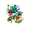
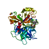
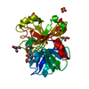
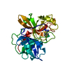
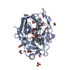
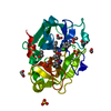
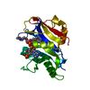
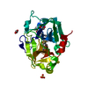
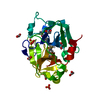
- PDB-7jsd: Hydroxylase homolog of BesD with Fe(II), alpha-ketoglutarate, and... -
+ Open data

- Basic information
| Entry | Database: PDB / ID: 7jsd | ||||||
|---|---|---|---|---|---|---|---|
| Title | Hydroxylase homolog of BesD with Fe(II), alpha-ketoglutarate, and lysine | ||||||
 Components Components | Lysine hydroxylase | ||||||
 Keywords Keywords | BIOSYNTHETIC PROTEIN / Hydroxylase / non-heme iron | ||||||
| Function / homology | q2cbj1_9rhob like domain / Jelly Rolls / Sandwich / Mainly Beta / 2-OXOGLUTARIC ACID / : / LYSINE Function and homology information Function and homology information | ||||||
| Biological species |  Streptomyces roseifaciens (bacteria) Streptomyces roseifaciens (bacteria) | ||||||
| Method |  X-RAY DIFFRACTION / SYNCHROTRON / MOLECULAR REPLACEMENT / Resolution: 2.5 Å X-RAY DIFFRACTION / SYNCHROTRON / MOLECULAR REPLACEMENT / Resolution: 2.5 Å | ||||||
 Authors Authors | Kissman, E.N. / Neugebauer, M.E. / Chang, M.C.Y. | ||||||
| Funding support |  United States, 1items United States, 1items
| ||||||
 Citation Citation | Journal: Nat.Chem.Biol. / Year: 2022 Title: Reaction pathway engineering converts a radical hydroxylase into a halogenase. Authors: Neugebauer, M.E. / Kissman, E.N. / Marchand, J.A. / Pelton, J.G. / Sambold, N.A. / Millar, D.C. / Chang, M.C.Y. | ||||||
| History |
|
- Structure visualization
| Structure viewer | Molecule: MolmilJmol/JSmol |
|---|
- Downloads & links
-Download
| PDBx/mmCIF format | 7jsd.cif.gz | 219.9 KB | Display | PDBx/mmCIF format |
|---|---|---|---|---|
| PDB format | pdb7jsd.ent.gz | 173.1 KB | Display | PDB format |
| PDBx/mmJSON format | 7jsd.json.gz | Tree view | PDBx/mmJSON format | |
| Others |  Other downloads Other downloads |
-Validation report
| Arichive directory | https://data.pdbj.org/pub/pdb/validation_reports/js/7jsdftp://data.pdbj.org/pub/pdb/validation_reports/js/7jsd | HTTPS FTP |
|---|
-Related structure data
| Related structure data |  6nieS S: Starting model for refinement |
|---|---|
| Similar structure data |

-Links
 PDBj
PDBj


- Assembly
| Deposited unit | 
| ||||||||
|---|---|---|---|---|---|---|---|---|---|
| 1 | 
| ||||||||
| 2 | 
| ||||||||
| 3 | 
| ||||||||
| 4 | 
| ||||||||
| Unit cell |
|
-Components
-Protein , 1 types, 4 molecules ABCD
| #1: Protein | Mass: 30078.697 Da / Num. of mol.: 4 Source method: isolated from a genetically manipulated source Source: (gene. exp.) Streptomyces roseifaciens (bacteria) / Production host: |
|---|
-Non-polymers , 5 types, 416 molecules 


| #2: Chemical |  Type: L-peptide linking / Mass: 147.195 Da / Num. of mol.: 3 / Source method: obtained synthetically / Formula: C6H15N2O2 / Feature type: SUBJECT OF INVESTIGATION Type: L-peptide linking / Mass: 147.195 Da / Num. of mol.: 3 / Source method: obtained synthetically / Formula: C6H15N2O2 / Feature type: SUBJECT OF INVESTIGATION#3: Chemical | ChemComp-AKG /  Mass: 146.098 Da / Num. of mol.: 4 / Source method: obtained synthetically / Formula: C5H6O5 / Feature type: SUBJECT OF INVESTIGATION Mass: 146.098 Da / Num. of mol.: 4 / Source method: obtained synthetically / Formula: C5H6O5 / Feature type: SUBJECT OF INVESTIGATION#4: Chemical | ChemComp-FE2 /  Mass: 55.845 Da / Num. of mol.: 4 / Source method: obtained synthetically / Formula: Fe / Feature type: SUBJECT OF INVESTIGATION Mass: 55.845 Da / Num. of mol.: 4 / Source method: obtained synthetically / Formula: Fe / Feature type: SUBJECT OF INVESTIGATION#5: Chemical |  Mass: 194.226 Da / Num. of mol.: 2 / Source method: obtained synthetically / Formula: C8H18O5 / Feature type: SUBJECT OF INVESTIGATION / Comment: precipitant*YM Mass: 194.226 Da / Num. of mol.: 2 / Source method: obtained synthetically / Formula: C8H18O5 / Feature type: SUBJECT OF INVESTIGATION / Comment: precipitant*YM#6: Water | ChemComp-HOH / | Mass: 18.015 Da / Num. of mol.: 403 / Source method: isolated from a natural source / Formula: H2O |
|---|
-Details
| Has ligand of interest | Y |
|---|
-Experimental details
-Experiment
| Experiment | Method: X-RAY DIFFRACTION / Number of used crystals: 1 |
|---|
- Sample preparation
| Crystal | Density Matthews: 2.58 Å3/Da / Density % sol: 52.34 % |
|---|---|
| Crystal grow | Temperature: 277 K / Method: vapor diffusion, hanging drop / pH: 7 Details: In an anaerobic chamber, equal volumes of protein solution (3 mg/mL Hydroxylase, lysine (3 mM), KG (3 mM, pH 7)) and reservoir solution (HEPES pH 7.0 (100 mM), sodium nitrate (200 mM) ...Details: In an anaerobic chamber, equal volumes of protein solution (3 mg/mL Hydroxylase, lysine (3 mM), KG (3 mM, pH 7)) and reservoir solution (HEPES pH 7.0 (100 mM), sodium nitrate (200 mM) containing 28% (w/v) PEG 3350); Crystals were soaked with iron chloride before flash freezing |
-Data collection
| Diffraction | Mean temperature: 194 K / Serial crystal experiment: N |
|---|---|
| Diffraction source | Source: SYNCHROTRON / Site: ALS / Beamline: 8.3.1 / Wavelength: 1.111 Å |
| Detector | Type: DECTRIS PILATUS3 S 6M / Detector: PIXEL / Date: Feb 15, 2020 |
| Radiation | Protocol: SINGLE WAVELENGTH / Monochromatic (M) / Laue (L): M / Scattering type: x-ray |
| Radiation wavelength | Wavelength: 1.111 Å / Relative weight: 1 |
| Reflection | Resolution: 2.5→87.081 Å / Num. obs: 40203 / % possible obs: 99.4 % / Redundancy: 6.9 % / Biso Wilson estimate: 38.69 Å2 / CC1/2: 0.987 / CC star: 0.997 / Rmerge(I) obs: 0.253 / Rrim(I) all: 0.2744 / Net I/σ(I): 6.2 |
| Reflection shell | Resolution: 2.5→2.589 Å / Rmerge(I) obs: 1.644 / Mean I/σ(I) obs: 1.42 / Num. unique obs: 3972 / CC1/2: 0.519 / CC star: 0.827 |
- Processing
| Software |
| ||||||||||||||||||||||||||||||||||||||||||||||||||||||||||||||||||||||||||||||||||||||||||
|---|---|---|---|---|---|---|---|---|---|---|---|---|---|---|---|---|---|---|---|---|---|---|---|---|---|---|---|---|---|---|---|---|---|---|---|---|---|---|---|---|---|---|---|---|---|---|---|---|---|---|---|---|---|---|---|---|---|---|---|---|---|---|---|---|---|---|---|---|---|---|---|---|---|---|---|---|---|---|---|---|---|---|---|---|---|---|---|---|---|---|---|
| Refinement | Method to determine structure: MOLECULAR REPLACEMENT Starting model: 6NIE Resolution: 2.5→87.08 Å / SU ML: 0.32 / Cross valid method: THROUGHOUT / σ(F): 1.34 / Phase error: 24.5 / Stereochemistry target values: ML
| ||||||||||||||||||||||||||||||||||||||||||||||||||||||||||||||||||||||||||||||||||||||||||
| Solvent computation | Shrinkage radii: 0.9 Å / VDW probe radii: 1.11 Å / Solvent model: FLAT BULK SOLVENT MODEL | ||||||||||||||||||||||||||||||||||||||||||||||||||||||||||||||||||||||||||||||||||||||||||
| Displacement parameters | Biso max: 111.85 Å2 / Biso mean: 43.7164 Å2 / Biso min: 13.89 Å2 | ||||||||||||||||||||||||||||||||||||||||||||||||||||||||||||||||||||||||||||||||||||||||||
| Refinement step | Cycle: final / Resolution: 2.5→87.08 Å
| ||||||||||||||||||||||||||||||||||||||||||||||||||||||||||||||||||||||||||||||||||||||||||
| Refine LS restraints |
| ||||||||||||||||||||||||||||||||||||||||||||||||||||||||||||||||||||||||||||||||||||||||||
| LS refinement shell | Refine-ID: X-RAY DIFFRACTION / Rfactor Rfree error: 0
|
