 ムービー
ムービー コントローラー


+ データを開く

- 基本情報
基本情報
| 登録情報 | データベース: PDB / ID: 7jmw | |||||||||
|---|---|---|---|---|---|---|---|---|---|---|
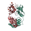
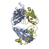
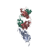
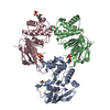
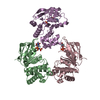
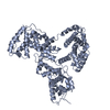
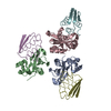
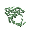
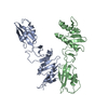
| タイトル | Crystal structure of SARS-CoV-2 spike protein receptor-binding domain in complex with cross-neutralizing antibody COVA1-16 Fab | |||||||||
 要素 要素 |
| |||||||||
 キーワード キーワード | VIRAL PROTEIN/IMMUNE SYSTEM / SARS-CoV-2 / Antibody / Spike / Coronavirus / COVID-19 / IMMUNE SYSTEM / VIRAL PROTEIN-IMMUNE SYSTEM complex | |||||||||
| 機能・相同性 |  機能・相同性情報 機能・相同性情報symbiont-mediated disruption of host tissue / Maturation of spike protein / Translation of Structural Proteins / Virion Assembly and Release / host cell surface / viral translation / host extracellular space / symbiont-mediated-mediated suppression of host tetherin activity / Induction of Cell-Cell Fusion / structural constituent of virion ...symbiont-mediated disruption of host tissue / Maturation of spike protein / Translation of Structural Proteins / Virion Assembly and Release / host cell surface / viral translation / host extracellular space / symbiont-mediated-mediated suppression of host tetherin activity / Induction of Cell-Cell Fusion / structural constituent of virion / membrane fusion / entry receptor-mediated virion attachment to host cell / Attachment and Entry / host cell endoplasmic reticulum-Golgi intermediate compartment membrane / positive regulation of viral entry into host cell / receptor-mediated virion attachment to host cell / host cell surface receptor binding / symbiont-mediated suppression of host innate immune response / receptor ligand activity / endocytosis involved in viral entry into host cell / fusion of virus membrane with host plasma membrane / fusion of virus membrane with host endosome membrane / viral envelope / symbiont entry into host cell / virion attachment to host cell / SARS-CoV-2 activates/modulates innate and adaptive immune responses / host cell plasma membrane / virion membrane / identical protein binding / membrane / plasma membrane 類似検索 - 分子機能 | |||||||||
| 生物種 |   Severe acute respiratory syndrome coronavirus 2 (ウイルス) Severe acute respiratory syndrome coronavirus 2 (ウイルス) Homo sapiens (ヒト) Homo sapiens (ヒト) | |||||||||
| 手法 |  X線回折 / シンクロトロン / 分子置換 / 解像度: 2.89 Å X線回折 / シンクロトロン / 分子置換 / 解像度: 2.89 Å | |||||||||
 データ登録者 データ登録者 | Liu, H. / Yuan, M. / Zhu, X. / Wu, N.C. / Wilson, I.A. | |||||||||
| 資金援助 |  米国, 2件 米国, 2件
| |||||||||
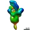
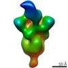
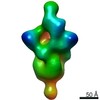
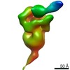
 引用 引用 | ジャーナル: Immunity / 年: 2020 タイトル: Cross-Neutralization of a SARS-CoV-2 Antibody to a Functionally Conserved Site Is Mediated by Avidity. 著者: Hejun Liu / Nicholas C Wu / Meng Yuan / Sandhya Bangaru / Jonathan L Torres / Tom G Caniels / Jelle van Schooten / Xueyong Zhu / Chang-Chun D Lee / Philip J M Brouwer / Marit J van Gils / ...著者: Hejun Liu / Nicholas C Wu / Meng Yuan / Sandhya Bangaru / Jonathan L Torres / Tom G Caniels / Jelle van Schooten / Xueyong Zhu / Chang-Chun D Lee / Philip J M Brouwer / Marit J van Gils / Rogier W Sanders / Andrew B Ward / Ian A Wilson /  要旨: Most antibodies isolated from individuals with coronavirus disease 2019 (COVID-19) are specific to severe acute respiratory syndrome coronavirus 2 (SARS-CoV-2). However, COVA1-16 is a relatively rare ...Most antibodies isolated from individuals with coronavirus disease 2019 (COVID-19) are specific to severe acute respiratory syndrome coronavirus 2 (SARS-CoV-2). However, COVA1-16 is a relatively rare antibody that also cross-neutralizes SARS-CoV. Here, we determined a crystal structure of the COVA1-16 antibody fragment (Fab) with the SARS-CoV-2 receptor-binding domain (RBD) and negative-stain electron microscopy reconstructions with the spike glycoprotein trimer to elucidate the structural basis of its cross-reactivity. COVA1-16 binds a highly conserved epitope on the SARS-CoV-2 RBD, mainly through a long complementarity-determining region (CDR) H3, and competes with the angiotensin-converting enzyme 2 (ACE2) receptor because of steric hindrance rather than epitope overlap. COVA1-16 binds to a flexible up conformation of the RBD on the spike and relies on antibody avidity for neutralization. These findings, along with the structural and functional rationale for epitope conservation, provide insights for development of more universal SARS-like coronavirus vaccines and therapies. #1: ジャーナル: Biorxiv / 年: 2020 タイトル: Cross-neutralization of a SARS-CoV-2 antibody to a functionally conserved site is mediated by avidity. 著者: Liu, H. / Wu, N.C. / Yuan, M. / Bangaru, S. / Torres, J.L. / Caniels, T.G. / van Schooten, J. / Zhu, X. / Lee, C.D. / Brouwer, P.J.M. / van Gils, M.J. / Sanders, R.W. / Ward, A.B. / Wilson, I.A. #2: ジャーナル: Immunity / 年: 2020タイトル: Cross-Neutralization of a SARS-CoV-2 Antibody to a Functionally Conserved Site Is Mediated by Avidity. 著者: Hejun Liu / Nicholas C Wu / Meng Yuan / Sandhya Bangaru / Jonathan L Torres / Tom G Caniels / Jelle van Schooten / Xueyong Zhu / Chang-Chun D Lee / Philip J M Brouwer / Marit J van Gils / ...著者: Hejun Liu / Nicholas C Wu / Meng Yuan / Sandhya Bangaru / Jonathan L Torres / Tom G Caniels / Jelle van Schooten / Xueyong Zhu / Chang-Chun D Lee / Philip J M Brouwer / Marit J van Gils / Rogier W Sanders / Andrew B Ward / Ian A Wilson / 要旨: Most antibodies isolated from individuals with coronavirus disease 2019 (COVID-19) are specific to severe acute respiratory syndrome coronavirus 2 (SARS-CoV-2). However, COVA1-16 is a relatively rare ...Most antibodies isolated from individuals with coronavirus disease 2019 (COVID-19) are specific to severe acute respiratory syndrome coronavirus 2 (SARS-CoV-2). However, COVA1-16 is a relatively rare antibody that also cross-neutralizes SARS-CoV. Here, we determined a crystal structure of the COVA1-16 antibody fragment (Fab) with the SARS-CoV-2 receptor-binding domain (RBD) and negative-stain electron microscopy reconstructions with the spike glycoprotein trimer to elucidate the structural basis of its cross-reactivity. COVA1-16 binds a highly conserved epitope on the SARS-CoV-2 RBD, mainly through a long complementarity-determining region (CDR) H3, and competes with the angiotensin-converting enzyme 2 (ACE2) receptor because of steric hindrance rather than epitope overlap. COVA1-16 binds to a flexible up conformation of the RBD on the spike and relies on antibody avidity for neutralization. These findings, along with the structural and functional rationale for epitope conservation, provide insights for development of more universal SARS-like coronavirus vaccines and therapies. | |||||||||
| 履歴 |
|
- 構造の表示
構造の表示
| 構造ビューア | 分子: MolmilJmol/JSmol |
|---|
- ダウンロードとリンク
-ダウンロード
| PDBx/mmCIF形式 | 7jmw.cif.gz | 305.7 KB | 表示 | PDBx/mmCIF形式 |
|---|---|---|---|---|
| PDB形式 | pdb7jmw.ent.gz | 206.5 KB | 表示 | PDB形式 |
| PDBx/mmJSON形式 | 7jmw.json.gz | ツリー表示 | PDBx/mmJSON形式 | |
| その他 |  その他のダウンロード その他のダウンロード |
-検証レポート
| 文書・要旨 | 7jmw_validation.pdf.gz | 751.9 KB | 表示 | wwPDB検証レポート |
|---|---|---|---|---|
| 文書・詳細版 | 7jmw_full_validation.pdf.gz | 759.4 KB | 表示 | |
| XML形式データ | 7jmw_validation.xml.gz | 23.5 KB | 表示 | |
| CIF形式データ | 7jmw_validation.cif.gz | 32 KB | 表示 | |
| アーカイブディレクトリ | https://data.pdbj.org/pub/pdb/validation_reports/jm/7jmwftp://data.pdbj.org/pub/pdb/validation_reports/jm/7jmw | HTTPS FTP |
-関連構造データ


-リンク
 PDBj
PDBj


- 集合体
集合体
| 登録構造単位 | 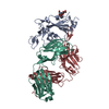
| ||||||||||||
|---|---|---|---|---|---|---|---|---|---|---|---|---|---|
| 1 | 
| ||||||||||||
| 単位格子 |
|
-要素
| #1: タンパク質 | 分子量: 26095.348 Da / 分子数: 1 / 由来タイプ: 組換発現 由来: (組換発現) Severe acute respiratory syndrome coronavirus 2 (ウイルス)遺伝子: S, 2 / 発現宿主:  Trichoplusia ni (イラクサキンウワバ) / 参照: UniProt: P0DTC2 Trichoplusia ni (イラクサキンウワバ) / 参照: UniProt: P0DTC2 |
|---|---|
| #2: 抗体 | 分子量: 25928.943 Da / 分子数: 1 / 由来タイプ: 組換発現 / 由来: (組換発現) Homo sapiens (ヒト) / 発現宿主:  |
| #3: 抗体 | 分子量: 23415.799 Da / 分子数: 1 / 由来タイプ: 組換発現 / 由来: (組換発現) Homo sapiens (ヒト) / 発現宿主: |
| #4: 多糖 | 2-acetamido-2-deoxy-beta-D-glucopyranose-(1-4)-2-acetamido-2-deoxy-beta-D-glucopyranose |
| 研究の焦点であるリガンドがあるか | Y |
| Has protein modification | Y |
-実験情報
-実験
| 実験 | 手法: X線回折 / 使用した結晶の数: 1 |
|---|
- 試料調製
試料調製
| 結晶 | マシュー密度: 2.72 Å3/Da / 溶媒含有率: 54.82 % |
|---|---|
| 結晶化 | 温度: 293.15 K / 手法: 蒸気拡散法, シッティングドロップ法 / pH: 6.9 / 詳細: 20% PEG 3350, 0.2 M Na-iodide, pH 6.9 |
-データ収集
| 回折 | 平均測定温度: 100 K / Serial crystal experiment: N |
|---|---|
| 放射光源 | 由来: シンクロトロン / サイト: SSRL / ビームライン: BL12-1 / 波長: 0.9795 Å |
| 検出器 | タイプ: DECTRIS EIGER X 16M / 検出器: PIXEL / 日付: 2020年6月10日 |
| 放射 | プロトコル: SINGLE WAVELENGTH / 単色(M)・ラウエ(L): M / 散乱光タイプ: x-ray |
| 放射波長 | 波長: 0.9795 Å / 相対比: 1 |
| 反射 | 解像度: 2.89→50 Å / Num. obs: 17656 / % possible obs: 97.9 % / 冗長度: 3.7 % / Biso Wilson estimate: 42.71 Å2 / CC1/2: 0.963 / Rpim(I) all: 0.09 / Rsym value: 0.153 / Net I/σ(I): 7.4 |
| 反射 シェル | 解像度: 2.89→2.95 Å / Mean I/σ(I) obs: 1.2 / Num. unique obs: 845 / CC1/2: 0.668 / Rpim(I) all: 0.429 / Rsym value: 0.691 |
- 解析
解析
| ソフトウェア |
| ||||||||||||||||||||||||||||||||||||||||||||||||||||||||
|---|---|---|---|---|---|---|---|---|---|---|---|---|---|---|---|---|---|---|---|---|---|---|---|---|---|---|---|---|---|---|---|---|---|---|---|---|---|---|---|---|---|---|---|---|---|---|---|---|---|---|---|---|---|---|---|---|---|
| 精密化 | 構造決定の手法: 分子置換 開始モデル: 6XC4,4IMK,2Q20 解像度: 2.89→42.77 Å / SU ML: 0.4316 / 交差検証法: FREE R-VALUE / σ(F): 1.35 / 位相誤差: 30.5748 立体化学のターゲット値: GeoStd + Monomer Library + CDL v1.2
| ||||||||||||||||||||||||||||||||||||||||||||||||||||||||
| 溶媒の処理 | 減衰半径: 0.9 Å / VDWプローブ半径: 1.11 Å / 溶媒モデル: FLAT BULK SOLVENT MODEL | ||||||||||||||||||||||||||||||||||||||||||||||||||||||||
| 原子変位パラメータ | Biso mean: 47.36 Å2 | ||||||||||||||||||||||||||||||||||||||||||||||||||||||||
| 精密化ステップ | サイクル: LAST / 解像度: 2.89→42.77 Å
| ||||||||||||||||||||||||||||||||||||||||||||||||||||||||
| 拘束条件 |
| ||||||||||||||||||||||||||||||||||||||||||||||||||||||||
| LS精密化 シェル |
| ||||||||||||||||||||||||||||||||||||||||||||||||||||||||
| 精密化 TLS | 手法: refined / Origin x: 32.6080084968 Å / Origin y: -24.9837704453 Å / Origin z: 36.0594463637 Å
| ||||||||||||||||||||||||||||||||||||||||||||||||||||||||
| 精密化 TLSグループ | Selection details: all |
