 Movie
Movie Controller

[English] 日本語
 Yorodumi
- PDB-2q20: Structure of the germline Vk1 O18/O8 light chain variable domain ... -
+ Open data

- Basic information
| Entry | Database: PDB / ID: 2q20 | ||||||
|---|---|---|---|---|---|---|---|
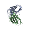
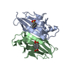
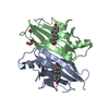
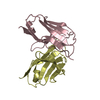
| Title | Structure of the germline Vk1 O18/O8 light chain variable domain homodimer | ||||||
 Components Components | Vk1 O18/O8 germline light chain variable domain | ||||||
 Keywords Keywords | PROTEIN FIBRIL / Vk1 / germline / AL / light chain amyloidosis / amyloid / immunoglobulin / light chain / light chain variable domain | ||||||
| Function / homology |  Function and homology information Function and homology informationCD22 mediated BCR regulation / Fc epsilon receptor (FCERI) signaling / Classical antibody-mediated complement activation / Initial triggering of complement / FCGR activation / Role of LAT2/NTAL/LAB on calcium mobilization / Role of phospholipids in phagocytosis / immunoglobulin complex / Scavenging of heme from plasma / antigen binding ...CD22 mediated BCR regulation / Fc epsilon receptor (FCERI) signaling / Classical antibody-mediated complement activation / Initial triggering of complement / FCGR activation / Role of LAT2/NTAL/LAB on calcium mobilization / Role of phospholipids in phagocytosis / immunoglobulin complex / Scavenging of heme from plasma / antigen binding / FCERI mediated Ca+2 mobilization / FCGR3A-mediated IL10 synthesis / Regulation of Complement cascade / Antigen activates B Cell Receptor (BCR) leading to generation of second messengers / Cell surface interactions at the vascular wall / FCGR3A-mediated phagocytosis / FCERI mediated MAPK activation / Regulation of actin dynamics for phagocytic cup formation / FCERI mediated NF-kB activation / Immunoregulatory interactions between a Lymphoid and a non-Lymphoid cell / blood microparticle / Potential therapeutics for SARS / adaptive immune response / immune response / extracellular region / identical protein binding / plasma membrane Similarity search - Function | ||||||
| Biological species |  Homo sapiens (human) Homo sapiens (human) | ||||||
| Method |  X-RAY DIFFRACTION / SYNCHROTRON / MOLECULAR REPLACEMENT / Resolution: 1.3 Å X-RAY DIFFRACTION / SYNCHROTRON / MOLECULAR REPLACEMENT / Resolution: 1.3 Å | ||||||
 Authors Authors | Thompson, J.R. / Ramirez-Alvarado, M. / Baden, E.M. | ||||||
 Citation Citation | Journal: J.Biol.Chem. / Year: 2008 Title: Altered dimer interface decreases stability in an amyloidogenic protein. Authors: Baden, E.M. / Owen, B.A. / Peterson, F.C. / Volkman, B.F. / Ramirez-Alvarado, M. / Thompson, J.R. | ||||||
| History |
| ||||||
| Remark 999 | Sequence The sequence is not available in the UniProt database at the time of processing. |
- Structure visualization
| Structure viewer | Molecule: MolmilJmol/JSmol |
|---|
- Downloads & links
-Download
| PDBx/mmCIF format | 2q20.cif.gz | 126.7 KB | Display | PDBx/mmCIF format |
|---|---|---|---|---|
| PDB format | pdb2q20.ent.gz | 101.4 KB | Display | PDB format |
| PDBx/mmJSON format | 2q20.json.gz | Tree view | PDBx/mmJSON format | |
| Others |  Other downloads Other downloads |
-Validation report
| Arichive directory | https://data.pdbj.org/pub/pdb/validation_reports/q2/2q20ftp://data.pdbj.org/pub/pdb/validation_reports/q2/2q20 | HTTPS FTP |
|---|
-Related structure data
| Related structure data |  2q1eC  1breS C: citing same article ( S: Starting model for refinement |
|---|---|
| Similar structure data |


-Links
 PDBj
PDBj


- Assembly
| Deposited unit | 
| ||||||||
|---|---|---|---|---|---|---|---|---|---|
| 1 |
| ||||||||
| Unit cell |
|
-Components
| #1: Antibody | Mass: 11971.120 Da / Num. of mol.: 2 Source method: isolated from a genetically manipulated source Source: (gene. exp.) Homo sapiens (human) / Gene: Vk1 O18/O8 light chain germline / Plasmid: pET12a / Production host:  #2: Water | ChemComp-HOH / |  Mass: 18.015 Da / Num. of mol.: 454 / Source method: isolated from a natural source / Formula: H2O Mass: 18.015 Da / Num. of mol.: 454 / Source method: isolated from a natural source / Formula: H2OHas protein modification | Y | |
|---|
-Experimental details
-Experiment
| Experiment | Method: X-RAY DIFFRACTION / Number of used crystals: 1 |
|---|
- Sample preparation
| Crystal | Density Matthews: 3.29 Å3/Da / Density % sol: 62.63 % |
|---|---|
| Crystal grow | Temperature: 295 K / Method: vapor diffusion, hanging drop Details: Protein: 890 micromolar in 10 mM TRIS pH 7.4. Mother Liquor: 15-30% (w/v) PEG 4K, 0.2 Li2SO4, 0.1 M TRIS pH 7.9-8.9, VAPOR DIFFUSION, HANGING DROP, temperature 295K |
-Data collection
| Diffraction | Mean temperature: 100 K |
|---|---|
| Diffraction source | Source: SYNCHROTRON / Site: APS  / Beamline: 19-BM / Wavelength: 1.0718 Å / Beamline: 19-BM / Wavelength: 1.0718 Å |
| Detector | Type: SBC-3 / Detector: CCD / Date: Feb 7, 2007 / Details: mirror |
| Radiation | Monochromator: Si111 / Protocol: SINGLE WAVELENGTH / Monochromatic (M) / Laue (L): M / Scattering type: x-ray |
| Radiation wavelength | Wavelength: 1.0718 Å / Relative weight: 1 |
| Reflection | Resolution: 1.3→99 Å / Num. all: 74839 / Num. obs: 74839 / % possible obs: 99.7 % / Redundancy: 12.2 % / Biso Wilson estimate: 22.2 Å2 / Rsym value: 0.046 / Net I/σ(I): 53.2 |
| Reflection shell | Resolution: 1.3→1.33 Å / Redundancy: 5.6 % / Mean I/σ(I) obs: 2.3 / Num. unique all: 5311 / Rsym value: 0.53 / % possible all: 96.3 |
- Processing
| Software |
| ||||||||||||||||||||||||||||||||||||||||||||||||||||||||||||||||||||||||||||||||||||||||||||||||||||||||||||||||||||||||||||||||||||||||||||||||||||||||||||||||||||||||||
|---|---|---|---|---|---|---|---|---|---|---|---|---|---|---|---|---|---|---|---|---|---|---|---|---|---|---|---|---|---|---|---|---|---|---|---|---|---|---|---|---|---|---|---|---|---|---|---|---|---|---|---|---|---|---|---|---|---|---|---|---|---|---|---|---|---|---|---|---|---|---|---|---|---|---|---|---|---|---|---|---|---|---|---|---|---|---|---|---|---|---|---|---|---|---|---|---|---|---|---|---|---|---|---|---|---|---|---|---|---|---|---|---|---|---|---|---|---|---|---|---|---|---|---|---|---|---|---|---|---|---|---|---|---|---|---|---|---|---|---|---|---|---|---|---|---|---|---|---|---|---|---|---|---|---|---|---|---|---|---|---|---|---|---|---|---|---|---|---|---|---|---|
| Refinement | Method to determine structure: MOLECULAR REPLACEMENT Starting model: polyserine model of pdb entry 1BRE chain A Resolution: 1.3→99 Å / Cor.coef. Fo:Fc: 0.984 / Cor.coef. Fo:Fc free: 0.977 / SU B: 0.995 / SU ML: 0.019 Isotropic thermal model: Isotropic model later refinement cycles anisotropic Cross valid method: THROUGHOUT / ESU R: 0.033 / ESU R Free: 0.035 / Stereochemistry target values: MAXIMUM LIKELIHOOD / Details: HYDROGENS HAVE BEEN ADDED IN THE RIDING POSITIONS
| ||||||||||||||||||||||||||||||||||||||||||||||||||||||||||||||||||||||||||||||||||||||||||||||||||||||||||||||||||||||||||||||||||||||||||||||||||||||||||||||||||||||||||
| Solvent computation | Ion probe radii: 0.8 Å / Shrinkage radii: 0.8 Å / VDW probe radii: 1.2 Å / Solvent model: BABINET MODEL WITH MASK | ||||||||||||||||||||||||||||||||||||||||||||||||||||||||||||||||||||||||||||||||||||||||||||||||||||||||||||||||||||||||||||||||||||||||||||||||||||||||||||||||||||||||||
| Displacement parameters | Biso mean: 14.491 Å2
| ||||||||||||||||||||||||||||||||||||||||||||||||||||||||||||||||||||||||||||||||||||||||||||||||||||||||||||||||||||||||||||||||||||||||||||||||||||||||||||||||||||||||||
| Refinement step | Cycle: LAST / Resolution: 1.3→99 Å
| ||||||||||||||||||||||||||||||||||||||||||||||||||||||||||||||||||||||||||||||||||||||||||||||||||||||||||||||||||||||||||||||||||||||||||||||||||||||||||||||||||||||||||
| Refine LS restraints |
| ||||||||||||||||||||||||||||||||||||||||||||||||||||||||||||||||||||||||||||||||||||||||||||||||||||||||||||||||||||||||||||||||||||||||||||||||||||||||||||||||||||||||||
| LS refinement shell | Resolution: 1.3→1.333 Å / Total num. of bins used: 20
|
