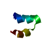
 Movie
Movie Controller


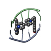
+ Open data

- Basic information
| Entry | Database: PDB / ID: 7jlh | ||||||
|---|---|---|---|---|---|---|---|
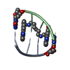
| Title | Structure of Centromeric Satellite III Non-canonical Duplex | ||||||
 Components Components | DNA AATGG | ||||||
 Keywords Keywords | DNA / non-canonical DNA / duplex / fiber / Satellite III | ||||||
| Function / homology | ACETATE ION / DNA Function and homology information Function and homology information | ||||||
| Biological species |  Homo sapiens (human) Homo sapiens (human) | ||||||
| Method |  X-RAY DIFFRACTION / SYNCHROTRON / MOLECULAR REPLACEMENT / Resolution: 1.57 Å X-RAY DIFFRACTION / SYNCHROTRON / MOLECULAR REPLACEMENT / Resolution: 1.57 Å | ||||||
 Authors Authors | Yatsunyk, L.A. / Chen, E.V. | ||||||
| Funding support |  United States, 1items United States, 1items
| ||||||
 Citation Citation | Journal: To Be Published Title: Crystal Structure of 4-repeat Satellite III Sequence with Non-Canonical Base Interactions Authors: Yatsunyk, L.A. / Chen, E.V. / Lee, H. | ||||||
| History |
|
- Structure visualization
| Structure viewer | Molecule: MolmilJmol/JSmol |
|---|
- Downloads & links
-Download
| PDBx/mmCIF format | 7jlh.cif.gz | 55.9 KB | Display | PDBx/mmCIF format |
|---|---|---|---|---|
| PDB format | pdb7jlh.ent.gz | 35 KB | Display | PDB format |
| PDBx/mmJSON format | 7jlh.json.gz | Tree view | PDBx/mmJSON format | |
| Others |  Other downloads Other downloads |
-Validation report
| Summary document | 7jlh_validation.pdf.gz | 406 KB | Display | wwPDB validaton report |
|---|---|---|---|---|
| Full document | 7jlh_full_validation.pdf.gz | 407.3 KB | Display | |
| Data in XML | 7jlh_validation.xml.gz | 4.4 KB | Display | |
| Data in CIF | 7jlh_validation.cif.gz | 5.8 KB | Display | |
| Arichive directory | https://data.pdbj.org/pub/pdb/validation_reports/jl/7jlhftp://data.pdbj.org/pub/pdb/validation_reports/jl/7jlh | HTTPS FTP |
-Related structure data
| Related structure data | 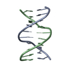 175dS S: Starting model for refinement |
|---|---|
| Similar structure data |

-Links
 PDBj
PDBj

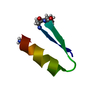
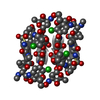
- Assembly
| Deposited unit | 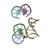
| ||||||||||||
|---|---|---|---|---|---|---|---|---|---|---|---|---|---|
| 1 | 
| ||||||||||||
| 2 | 
| ||||||||||||
| 3 | 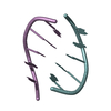
| ||||||||||||
| Unit cell |
|
-Components
| #1: DNA chain | Mass: 1544.061 Da / Num. of mol.: 6 / Source method: obtained synthetically Details: DNA consists of four ATGGA repeats. Packing of DNA into crystal structure leads to the presence of three antiparallel AATGG duplexes in the asymmetric unit. Source: (synth.) Homo sapiens (human)#2: Chemical | ChemComp-ACT / |   Mass: 59.044 Da / Num. of mol.: 1 / Source method: obtained synthetically / Formula: C2H3O2 Mass: 59.044 Da / Num. of mol.: 1 / Source method: obtained synthetically / Formula: C2H3O2#3: Chemical | ChemComp-MG / |   Mass: 24.305 Da / Num. of mol.: 1 / Source method: obtained synthetically / Formula: Mg Mass: 24.305 Da / Num. of mol.: 1 / Source method: obtained synthetically / Formula: Mg#4: Water | ChemComp-HOH / |  Mass: 18.015 Da / Num. of mol.: 74 / Source method: isolated from a natural source / Formula: H2O Mass: 18.015 Da / Num. of mol.: 74 / Source method: isolated from a natural source / Formula: H2OHas ligand of interest | N | Sequence details | The full DNA sequence is d(ATGGA)4 which forms self complimentary duplex. Due to the repetitive ...The full DNA sequence is d(ATGGA)4 which forms self complimentary duplex. Due to the repetitive nature and symmetry of such duplex, the asymmetric unit only includes 1/4 of the duplex, with the sequences AATGG. There are three such duplexes in the ASU. | |
|---|
-Experimental details
-Experiment
| Experiment | Method: X-RAY DIFFRACTION / Number of used crystals: 1 |
|---|
- Sample preparation
| Crystal | Density Matthews: 2.19 Å3/Da / Density % sol: 48.22 % |
|---|---|
| Crystal grow | Temperature: 293 K / Method: vapor diffusion, hanging drop / pH: 6.5 Details: Tris-Acetate, potassium acetate, magnesium acetate, MES monohydrate, 2-Methyl-2,4-pentanediol |
-Data collection
| Diffraction | Mean temperature: 196 K / Serial crystal experiment: N |
|---|---|
| Diffraction source | Source: SYNCHROTRON / Site: APS / Beamline: 24-ID-E / Wavelength: 1.0711 Å |
| Detector | Type: DECTRIS EIGER X 16M / Detector: PIXEL / Date: Mar 23, 2019 |
| Radiation | Monochromator: Si(220) / Protocol: SINGLE WAVELENGTH / Monochromatic (M) / Laue (L): M / Scattering type: x-ray |
| Radiation wavelength | Wavelength: 1.0711 Å / Relative weight: 1 |
| Reflection | Resolution: 1.57→32.54 Å / Num. obs: 12855 / % possible obs: 99.7 % / Redundancy: 5.9 % / Biso Wilson estimate: 31.32 Å2 / CC1/2: 0.998 / Net I/σ(I): 15.1 |
| Reflection shell | Resolution: 1.57→1.63 Å / Redundancy: 5.1 % / Mean I/σ(I) obs: 2.5 / Num. unique obs: 643 / CC1/2: 0.991 / % possible all: 99 |
- Processing
| Software |
| ||||||||||||||||||||||||||||||||||||||||||||||||||||||||||||||||||||||
|---|---|---|---|---|---|---|---|---|---|---|---|---|---|---|---|---|---|---|---|---|---|---|---|---|---|---|---|---|---|---|---|---|---|---|---|---|---|---|---|---|---|---|---|---|---|---|---|---|---|---|---|---|---|---|---|---|---|---|---|---|---|---|---|---|---|---|---|---|---|---|---|
| Refinement | Method to determine structure: MOLECULAR REPLACEMENT Starting model: 175D Resolution: 1.57→32.54 Å / SU ML: 0.2088 / Cross valid method: FREE R-VALUE / σ(F): 1.35 / Phase error: 32.2896 Stereochemistry target values: GeoStd + Monomer Library + CDL v1.2
| ||||||||||||||||||||||||||||||||||||||||||||||||||||||||||||||||||||||
| Solvent computation | Shrinkage radii: 0.9 Å / VDW probe radii: 1.11 Å / Solvent model: FLAT BULK SOLVENT MODEL | ||||||||||||||||||||||||||||||||||||||||||||||||||||||||||||||||||||||
| Displacement parameters | Biso mean: 48.43 Å2 | ||||||||||||||||||||||||||||||||||||||||||||||||||||||||||||||||||||||
| Refinement step | Cycle: LAST / Resolution: 1.57→32.54 Å
| ||||||||||||||||||||||||||||||||||||||||||||||||||||||||||||||||||||||
| Refine LS restraints |
| ||||||||||||||||||||||||||||||||||||||||||||||||||||||||||||||||||||||
| LS refinement shell |
| ||||||||||||||||||||||||||||||||||||||||||||||||||||||||||||||||||||||
| Refinement TLS params. | Method: refined / Origin x: 0.832115918806 Å / Origin y: -19.1431532399 Å / Origin z: -3.07229969381 Å
| ||||||||||||||||||||||||||||||||||||||||||||||||||||||||||||||||||||||
| Refinement TLS group | Selection details: all |
