 Movie
Movie Controller


+ Open data

- Basic information
| Entry | Database: PDB / ID: 1hhz | |||||||||
|---|---|---|---|---|---|---|---|---|---|---|
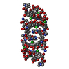
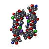
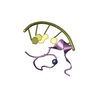
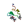
| Title | Deglucobalhimycin in complex with cell wall pentapeptide | |||||||||
 Components Components |
| |||||||||
 Keywords Keywords | ANTIBIOTIC/PEPTIDE / ANTIBIOTIC-PEPTIDE COMPLEX / GLYCOPEPTIDE / ANTIBIOTIC / CELL WALL PEPTIDE / BALHIMYCIN | |||||||||
| Function / homology | Deglucobalhimycin / Chem-DVC / R-1,2-PROPANEDIOL / :  Function and homology information Function and homology information | |||||||||
| Biological species |  AMYCOLATOPSIS SP. (bacteria) AMYCOLATOPSIS SP. (bacteria)synthetic construct (others) | |||||||||
| Method |  X-RAY DIFFRACTION / SYNCHROTRON / DIRECT METHODS / Resolution: 0.99 Å X-RAY DIFFRACTION / SYNCHROTRON / DIRECT METHODS / Resolution: 0.99 Å | |||||||||
 Authors Authors | Lehmann, C. / Bunkoczi, G. / Sheldrick, G.M. / Vertesy, L. | |||||||||
 Citation Citation | Journal: J.Mol.Biol. / Year: 2002 Title: Structures of Glycopeptide Antibiotics with Peptides that Model Bacterial Cell-Wall Precursors Authors: Lehmann, C. / Bunkoczi, G. / Vertesy, L. / Sheldrick, G.M. | |||||||||
| History |
|
- Structure visualization
| Structure viewer | Molecule: MolmilJmol/JSmol |
|---|
- Downloads & links
-Download
| PDBx/mmCIF format | 1hhz.cif.gz | 35.9 KB | Display | PDBx/mmCIF format |
|---|---|---|---|---|
| PDB format | pdb1hhz.ent.gz | 27.4 KB | Display | PDB format |
| PDBx/mmJSON format | 1hhz.json.gz | Tree view | PDBx/mmJSON format | |
| Others |  Other downloads Other downloads |
-Validation report
| Arichive directory | https://data.pdbj.org/pub/pdb/validation_reports/hh/1hhzftp://data.pdbj.org/pub/pdb/validation_reports/hh/1hhz | HTTPS FTP |
|---|
-Related structure data


-Links
 PDBj
PDBj- Assembly
| Deposited unit | 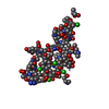
| ||||||||||||
|---|---|---|---|---|---|---|---|---|---|---|---|---|---|
| 1 | 
| ||||||||||||
| 2 | 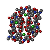
| ||||||||||||
| Unit cell |
| ||||||||||||
| Components on special symmetry positions |
| ||||||||||||
| Noncrystallographic symmetry (NCS) | NCS oper:
|
-Components
| #1: Protein/peptide | 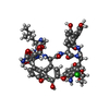  Type: Glycopeptide / Class: Antibiotic, Antimicrobial / Mass: 1149.977 Da / Num. of mol.: 3 / Source method: obtained synthetically Type: Glycopeptide / Class: Antibiotic, Antimicrobial / Mass: 1149.977 Da / Num. of mol.: 3 / Source method: obtained syntheticallyDetails: DEGLUCOBALHIMYCIN LACKS THE D-GLUCOSE COMPONENT OF BALHIMYCIN CONSISTING OF THE TRICYCLIC HEPTAPEPTIDE AND (2R,4S,6S)-4-AZANYL-4,6-DIMETHYL-OXANE-2,5,5-TRIOL ONLY LINKED TO RESIDUE 6. Source: (synth.) AMYCOLATOPSIS SP. (bacteria) / References: NOR: NOR00707, Deglucobalhimycin#2: Protein/peptide | Mass: 489.542 Da / Num. of mol.: 3 / Source method: obtained synthetically / Source: (synth.) synthetic construct (others) #3: Sugar |   Type: L-saccharide, alpha linking, Glycopeptide / Class: Antibiotic, Antimicrobial / Mass: 177.198 Da / Num. of mol.: 3 Type: L-saccharide, alpha linking, Glycopeptide / Class: Antibiotic, Antimicrobial / Mass: 177.198 Da / Num. of mol.: 3Source method: isolated from a genetically manipulated source Formula: C7H15NO4 Details: DEGLUCOBALHIMYCIN LACKS THE D-GLUCOSE COMPONENT OF BALHIMYCIN CONSISTING OF THE TRICYCLIC HEPTAPEPTIDE AND (2R,4S,6S)-4-AZANYL-4,6-DIMETHYL-OXANE-2,5,5-TRIOL ONLY LINKED TO RESIDUE 6. References: Deglucobalhimycin #4: Chemical | ChemComp-PGR /   Mass: 76.094 Da / Num. of mol.: 4 / Source method: obtained synthetically / Formula: C3H8O2 Mass: 76.094 Da / Num. of mol.: 4 / Source method: obtained synthetically / Formula: C3H8O2#5: Water | ChemComp-HOH / |  Mass: 18.015 Da / Num. of mol.: 95 / Source method: isolated from a natural source / Formula: H2O Mass: 18.015 Da / Num. of mol.: 95 / Source method: isolated from a natural source / Formula: H2OCompound details | BALHIMYCIN IS A TRICYCLIC GLYCOPEPTIDE. THE SCAFFOLD IS A HEPTAPEPTIDE WITH THE CONFIGURATION D-D-L- ...BALHIMYCIN | Has protein modification | Y | |
|---|
-Experimental details
-Experiment
| Experiment | Method: X-RAY DIFFRACTION / Number of used crystals: 1 |
|---|
- Sample preparation
| Crystal | Density Matthews: 2.7 Å3/Da / Density % sol: 38.1 % |
|---|---|
| Crystal grow | pH: 7 / Details: 0.3M CIT, PH=7, 30% 1,2-PROPANEDIOL, pH 7.00 |
-Data collection
| Diffraction | Mean temperature: 100 K |
|---|---|
| Diffraction source | Source: SYNCHROTRON / Site: EMBL/DESY, HAMBURG  / Beamline: X11 / Wavelength: 0.9114 / Beamline: X11 / Wavelength: 0.9114 |
| Detector | Type: MARRESEARCH / Detector: IMAGE PLATE / Date: Mar 15, 1999 |
| Radiation | Protocol: SINGLE WAVELENGTH / Monochromatic (M) / Laue (L): M / Scattering type: x-ray |
| Radiation wavelength | Wavelength: 0.9114 Å / Relative weight: 1 |
| Reflection | Resolution: 0.99→41.85 Å / Num. obs: 29932 / % possible obs: 99.9 % / Redundancy: 7.5 % / Rmerge(I) obs: 0.053 |
| Reflection shell | Resolution: 0.99→1.1 Å / Redundancy: 7.5 % / Rmerge(I) obs: 0.19 / % possible all: 100 |
- Processing
| Software |
| |||||||||||||||||||||||||||||||||
|---|---|---|---|---|---|---|---|---|---|---|---|---|---|---|---|---|---|---|---|---|---|---|---|---|---|---|---|---|---|---|---|---|---|---|
| Refinement | Method to determine structure: DIRECT METHODS / Resolution: 0.99→41.85 Å / Num. parameters: 4237 / Num. restraintsaints: 4905 / Cross valid method: THROUGHOUT / Stereochemistry target values: ENGH AND HUBER
| |||||||||||||||||||||||||||||||||
| Solvent computation | Solvent model: METHOD USED: MOEWS & KRETSINGER, J.MOL.BIOL.91(1973)201-228 | |||||||||||||||||||||||||||||||||
| Refine analyze | Num. disordered residues: 6 / Occupancy sum hydrogen: 280.8 / Occupancy sum non hydrogen: 448.5 | |||||||||||||||||||||||||||||||||
| Refinement step | Cycle: LAST / Resolution: 0.99→41.85 Å
| |||||||||||||||||||||||||||||||||
| Refine LS restraints |
|
