Mean temperature: 100 K / Serial crystal experiment: N
Diffraction source
Source: SYNCHROTRON / Site: ALS / Beamline: 4.2.2 / Wavelength: 1.00003 Å
Detector
Type: NOIR-1 / Detector: CCD / Date: Jan 30, 2015
Radiation
Protocol: SINGLE WAVELENGTH / Monochromatic (M) / Laue (L): M / Scattering type: x-ray
Radiation wavelength
Wavelength: 1.00003 Å / Relative weight: 1
Reflection
Resolution: 2.1→51.49 Å / Num. obs: 10615 / % possible obs: 94 % / Redundancy: 10.8 % / Biso Wilson estimate: 25.18 Å2 / Rrim(I) all: 0.2348 / Net I/σ(I): 3.28
Reflection shell
Resolution: 2.1→51.49 Å / Num. unique obs: 1041 / Rrim(I) all: 0.2348 / % possible all: 94.13
-
Processing
Software
Name
Version
Classification
REFMAC
5.8.0258
refinement
HKL-2000
datareduction
HKL-2000
datascaling
PHASER
phasing
Refinement
Method to determine structure: AB INITIO PHASING / Resolution: 2.1→51.49 Å / Cor.coef. Fo:Fc: 0.955 / Cor.coef. Fo:Fc free: 0.924 / SU B: 6.369 / SU ML: 0.16 / Cross valid method: THROUGHOUT / ESU R: 0.251 / ESU R Free: 0.196 / Stereochemistry target values: MAXIMUM LIKELIHOOD Details: HYDROGENS HAVE BEEN ADDED IN THE RIDING POSITIONS U VALUES : REFINED INDIVIDUALLY
Rfactor
Num. reflection
% reflection
Selection details
Rfree
0.23437
592
5.3 %
RANDOM
Rwork
0.19211
-
-
-
obs
0.19444
10615
94.2 %
-
Solvent computation
Ion probe radii: 0.9 Å / Shrinkage radii: 0.9 Å / VDW probe radii: 1.2 Å / Solvent model: MASK
Displacement parameters
Biso mean: 33.401 Å2
Baniso -1
Baniso -2
Baniso -3
1-
4.99 Å2
0 Å2
0.4 Å2
2-
-
-1.64 Å2
-0 Å2
3-
-
-
-3.36 Å2
Refinement step
Cycle: LAST / Resolution: 2.1→51.49 Å
Protein
Nucleic acid
Ligand
Solvent
Total
Num. atoms
1535
0
0
27
1562
Refine LS restraints
Refine-ID
Type
Dev ideal
Dev ideal target
Number
X-RAY DIFFRACTION
r_bond_refined_d
0.008
0.019
1544
X-RAY DIFFRACTION
r_bond_other_d
0.001
0.02
1464
X-RAY DIFFRACTION
r_angle_refined_deg
1.305
1.902
2069
X-RAY DIFFRACTION
r_angle_other_deg
1.015
2.934
3401
X-RAY DIFFRACTION
r_dihedral_angle_1_deg
5.85
5
192
X-RAY DIFFRACTION
r_dihedral_angle_2_deg
35.061
25.769
78
X-RAY DIFFRACTION
r_dihedral_angle_3_deg
13.67
15
314
X-RAY DIFFRACTION
r_dihedral_angle_4_deg
22.748
15
11
X-RAY DIFFRACTION
r_chiral_restr
0.077
0.2
243
X-RAY DIFFRACTION
r_gen_planes_refined
0.004
0.02
1705
X-RAY DIFFRACTION
r_gen_planes_other
0.001
0.02
286
X-RAY DIFFRACTION
r_mcbond_it
3.602
3.099
774
X-RAY DIFFRACTION
r_mcbond_other
3.585
3.095
773
X-RAY DIFFRACTION
r_mcangle_it
5.112
4.629
964
X-RAY DIFFRACTION
r_mcangle_other
5.11
4.634
965
X-RAY DIFFRACTION
r_scbond_it
4.462
3.636
770
X-RAY DIFFRACTION
r_scbond_other
4.459
3.64
771
X-RAY DIFFRACTION
r_scangle_other
6.536
5.241
1106
X-RAY DIFFRACTION
r_long_range_B_refined
8.4
36.654
1615
X-RAY DIFFRACTION
r_long_range_B_other
8.401
36.691
1614
LS refinement shell
Resolution: 2.1→2.154 Å / Total num. of bins used: 20
Rfactor
Num. reflection
% reflection
Rfree
0.305
34
-
Rwork
0.258
720
-
obs
-
-
86.77 %
+
About Yorodumi
-
News
-
Feb 9, 2022. New format data for meta-information of EMDB entries
New format data for meta-information of EMDB entries
Version 3 of the EMDB header file is now the official format.
The previous official version 1.9 will be removed from the archive.
In the structure databanks used in Yorodumi, some data are registered as the other names, "COVID-19 virus" and "2019-nCoV". Here are the details of the virus and the list of structure data.
Jan 31, 2019. EMDB accession codes are about to change! (news from PDBe EMDB page)
EMDB accession codes are about to change! (news from PDBe EMDB page)
The allocation of 4 digits for EMDB accession codes will soon come to an end. Whilst these codes will remain in use, new EMDB accession codes will include an additional digit and will expand incrementally as the available range of codes is exhausted. The current 4-digit format prefixed with “EMD-” (i.e. EMD-XXXX) will advance to a 5-digit format (i.e. EMD-XXXXX), and so on. It is currently estimated that the 4-digit codes will be depleted around Spring 2019, at which point the 5-digit format will come into force.
The EM Navigator/Yorodumi systems omit the EMD- prefix.
Related info.:Q: What is EMD? / ID/Accession-code notation in Yorodumi/EM Navigator
Yorodumi is a browser for structure data from EMDB, PDB, SASBDB, etc.
This page is also the successor to EM Navigator detail page, and also detail information page/front-end page for Omokage search.
The word "yorodu" (or yorozu) is an old Japanese word meaning "ten thousand". "mi" (miru) is to see.
Related info.:EMDB / PDB / SASBDB / Comparison of 3 databanks / Yorodumi Search / Aug 31, 2016. New EM Navigator & Yorodumi / Yorodumi Papers / Jmol/JSmol / Function and homology information / Changes in new EM Navigator and Yorodumi
 Movie
Movie Controller

 Yorodumi
 Open data
 Basic information
 Components
 Keywords
 Function and homology information
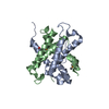
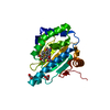
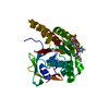
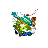
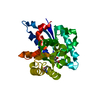
 Staphylococcus aureus (bacteria)
 X-RAY DIFFRACTION /
  Authors
 United States, 1items
  Citation
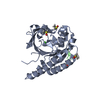
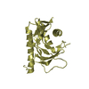
 Structure visualization
 Downloads & links
 Other downloads
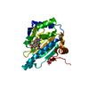


 PDBj
PDBj


 Assembly
 Mass: 18.015 Da / Num. of mol.: 27 / Source method: isolated from a natural source / Formula: H2O
 Sample preparation
 Processing